Astelin dosages: 10 ml
Astelin packs: 1 sprayer, 2 sprayer, 3 sprayer, 4 sprayer, 5 sprayer, 6 sprayer, 7 sprayer, 8 sprayer, 9 sprayer, 10 sprayer
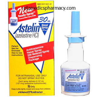
10 ml astelin discount with mastercard
Clinical lesions present purple brown painful oedema which can evolve into necrotic plaques that heal with sclerotic allergy testing joondalup astelin 10 ml generic, indurated scars which may turn into sure to underlying muscle and bone [25] allergy medicine and high blood pressure 10 ml astelin cheap overnight delivery. Similar findings have been described in the penis following a grease gun injury [42]. Exogenous oils may be highlighted by particular stains corresponding to oil red O and osmium tetroxide [2]. Histopathological findings in local reactions to implants of silicone are variable depending primarily on the form of the injected silicone. Solid elastomer silicone induces an exuberant foreign physique granulomatous response, whereas silicone oil and gel induce a sparser inflammatory response. Silicone particles seem as groups of spherical empty vacuoles of various sizes between collagen bundles or inside macrophages. Granulomas from collagenbased cosmetic fillers containing polymethylmethacrylate microspheres present a nodular or diffuse granulomatous infiltrate surrounding rounded vacuoles of similar form and measurement, which mimic normal adipocytes and correspond to the implanted microspheres [5]. Injections of lipomas with phosphatidylcholinecontaining substances induce an early response characterised by neutrophilic infiltration with partially destroyed fat cells; late lesions present infiltration of T lymphocytes and macrophages with foamy histiocytes, accompanied by thickened septa and pseudocapsule formation surrounding the inflamed space [9]. Persistent reactions to aluminium at the web site of injection of hyposensitization vaccines present abundant lymphoid follicles in the subcutaneous tissue with germinal centre formation. They may mimic lupus profundus, pseudolymphoma or deep morphoea, however the plentiful eosinophils and the identification of characteristic histiocytes with basophilic granular cytoplasm are the important thing distinctive options allowing the correct prognosis [19]. These macrophages comprise lysosomes crammed with aluminium salts that can be demonstrated with Xray dispersion microanalysis. Pentazozine panniculitis is manifested as sclerodermoid plaques that end result from thrombosis of small vessels, endarteritis, granulomatous inflammation, lipophagic granulomata and pronounced fibrosis of the dermis and subcutaneous fats [11,44]. Panniculitis secondary to vitamin K injections is also characterised by distinguished sclerosis of the collagen bundles of the connective tissue septa of the subcutis and an inflammatory infiltrate of lymphocytes, mast cells and plasma cells, which raises the histopathological differential analysis with morphoea [16,38]. In distinction with deep morphoea, vitamin K1 panniculitis often additionally involves the fat lobule with lipophagic granulomata. Povidone panniculitis reveals granulomatous infiltration of the fats lobule with focal haemorrhage and necrosis. Many macrophages contain greyblue foamy materials in their cytoplasm, which is optimistic for Congo purple and chlorazolfast pink [39]. Extravasation of cytotoxic drugs exhibits lobular panniculitis, abundant adipocyte necrosis with little inflammatory infiltrate together with epidermal lesions attributable to direct cytotoxicity. In distinction, the lymphoid follicles within the septa and on the interface between septum and fats lobule are mainly composed of B lymphocytes [17]. Factitious panniculitis due to repeated trauma exhibits organizing haematomas, focal granulomas and haemosiderin deposition [29]. If artefact is suspected, the affected space could additionally be occluded for per week with a bandage: improvement would support a suspicion of self induced factitious panniculitis, for which acceptable social and psychiatric care should be supplied. Panniculitis secondary to cosmetic fillers normally require intralesional steroids and, if attainable, removal of the implanted material. Panniculitis secondary to injection of medication normally requires only supportive care and withdrawal of the accountable drug. Neutrophilic lobular panniculitis definition Neutrophilic lobular panniculitis incorporates a range of different panniculitides during which the fats lobule infiltrate is mostly composed of neutrophils (Box ninety nine. Histopathologically, cutaneous lesions present oedema of the papillary dermis and a dense bandlike infiltrate of neutrophils involving largely the superficial dermis, with no vasculitis [3]. A prognosis of subcutaneous Sweet syndrome must be made, nevertheless, solely in those instances by which the neutrophilic infiltrate includes solely the subcutaneous tissue with few or no neutrophils in the dermis. Presentation Subcutaneous Sweet syndrome, like classical Sweet syndrome, has a median age of onset in the course of the sixth decade of life however appears to not present the feminine preponderance of the latter [3,8]. Frequently, the onset of subcutaneous nodules is preceded or accompanied by systemic symptoms corresponding to fever and malaise [4,9,eleven,13,15,16]; leukocytosis was present in several sufferers [11�13,15,16]. The most frequent places are the lower extremities [10�14,sixteen,17], followed by higher extremities [11,12,14], trunk [9,11,12,14] and head [9]. Investigations Histopathological study of the lesions demonstrates a dense infiltrate of mature neutrophils involving subcutaneous tissue. Vasculitis is normally absent in all cases, however in two sufferers leukocytoclasia was noted [9,12]. Occasionally, some mononuclear cells could also be discovered in the subcutaneous tissue [9,13]. Rarely, infiltration of myeloperoxidase positive immature granulocytes has been described [20], representing the subcutaneous counterpart of the socalled histiocytoid Sweet syndrome [29]. In summary, Sweet syndrome may involve subcutaneous tissue with two different patterns: (i) with a largely septal panniculitis and infrequently granulomatous infiltrate, as in classical erythema nodosum related to Sweet syndrome [30]; and (ii) with a neutrophilic infiltrate principally involving the fats lobules, as is the case in subcutaneous Sweet syndrome. Management Most sufferers with subcutaneous Sweet syndrome present dramatic response to systemic corticosteroids, such as prednisolone [9,10,13�15]. They include nodules on the lower extremities [32,33], and typically additionally on the arms [32,33], equivalent to these of classical erythema nodosum [31]. However, histopathological research demonstrates involvement of both septa and fat lobules [32,33], with necrotizing leukocytoclastic vasculitis involving arterioles and venules [31�34]. Most of the circumstances present a neutrophilic lobular panniculitis, though in rare situations a septal element may be predominant [11]. To date, only 22 well documented cases of subcutaneous Sweet syndrome have been reported [9�20,21,22�26]. Several features support the connection of neutrophilic lobular panniculitis to Sweet syndrome. In some circumstances, subcutaneous Sweet syndrome was adopted by classical dermal Sweet syndrome [4], whereas one other affected person presented simultaneously with classical Sweet syndrome and Sweet panniculitis [17]. As in classical Sweet syndrome, many sufferers with subcutaneous Sweet syndrome had associated myelodysplastic syndromes and haematological neoplasms [9,12�14,18,19,23,24,26�28], and cutaneous lesions showed a wonderful response to systemic corticosteroids. Fat necrosis has been present in nearly all cases [36,37,41,42] and leukocytoclastic vasculitis was described in some [36,42]. Bowelassociated dermatosisarthritis syndrome (see Chapter 152) the bowelassociated dermatosis�arthritis syndrome is characterised by recurrent fever, arthralgia and pores and skin lesions after intestinal bypass or bariatric surgical procedure [43,44]. The most frequent skin lesions include erythematous papules or vesiculopustules. Lobular neutrophilic panniculitis with tender subcutaneous nodules on the decrease extremities has, however, not often been described in these patients [43�46]. Subcutaneous sarcoidosis definition and nomenclature Minimal dermal involvement is appropriate for a histopathological analysis of subcutaneous sarcoidosis [1], however subcutaneous sarcoidosis is considered a selected clinicopathological variant of sarcoidosis involving completely the subcutaneous fats and ought to be differentiated from nodular dermal lesions of sarcoidosis with deep extension into the subcutaneous tissue [2]. Mostly, subcutaneous sarcoidosis is associated with systemic sarcoidosis, though with an indolent and nonaggressive form of the illness [3]. However, patients with systemic sarcoidosis may also develop sarcoidal granulomas involving the subcutaneous tissue as a particular cutaneous lesion and which is referred to as subcutaneous sarcoidosis. Subcutaneous sarcoidosis is a uncommon form of cutaneous sarcoidosis [3�6] (see Chapter 98). Subcutaneous sarcoidosis is the least common particular cutaneous manifestation of sarcoidosis. Rheumatoid arthritis (see Chapter 154) Several cases of neutrophilic (pustular) panniculitis have been described in patients with rheumatoid arthritis [35�41]. In these sufferers, subcutaneous nodules are principally located on the decrease extremities they usually present a bent to type draining fistulae with a yellowish discharge [36,40�42].
Syndromes
- Slowing of activity
- CK isoenzymes
- High or very low temperature, chills
- Headache
- Shock wave lithotripsy
- Fish or oysters
- Are both sides of your neck affected equally?
Astelin 10 ml generic
Clinical classification of itch: a Position Paper of the International Forum for the Study of Itch allergy symptoms ginger and hon astelin 10 ml order visa. The auricle is attached to the head by fibrous ligaments and three vestigial auricularis muscular tissues allergy treatment children cheap astelin 10 ml on line. The size and common element of the auricle can range greatly between individuals, and could additionally be characteristically affected in numerous congenital syndromes. The dermis of the ear has a fancy dermal�epidermal junction, a conspicuous stratum granulosum and a thick, compact stratum corneum. Sebaceous glands are numerous, particularly on the tragus and lobe, and fine vellus or terminal hairs occur over the whole surface, but are particularly distinguished on the helix and tragus. Eccrine sweat glands are sparsely and irregularly distributed except in the external auditory canal, which has, instead, a lot of modified apocrine or ceruminous glands. The pinna has a variably thick fatty layer that extends between the perichondrium and the reticular dermis and that also varieties the principle fibrofatty core of the lobe of the ear. The blood supply to the auricle is offered by anastomosing branches of the superficial temporal and posterior auricular arteries, which drain by way of posterior auricular and superficial temporal veins into the exterior jugular vein and through the superficial temporal, maxillary and facial veins into the interior jugular vein. Lymphatic drainage is to the superficial parotid, retroauricular and superficial cervical lymph nodes. The back of the ear is equipped by the larger auricular nerve (C2,3), the concha by the auricular branch of the vagus (Xth) and the anterior a part of the pinna and the external auditory canal by the auriculotemporal department of the Vth cranial nerve. With this difficult nerve supply, otalgia is extra generally as a outcome of referred ache than to illness in the ear itself [4]. Within the dermis, the nerve supply is abundant, especially round hair follicles the place there are complicated basketlike networks of acetylcholinesterase and butyrylcholinesterase nerve fibres. The exterior auditory canal extends upwards and backwards in an Sshaped curve from the concha to the tympanic membrane. The angle of curvature varies between races and people, being more marked in white folks than in black people or Polynesians. The outer third of the canal is cartilaginous and is lined by a thicker layer of skin than the internal portion inside the temporal bone. Anteroinferiorly there are two horizontal fissures in the cartilaginous canal, the fissures of Santorini. These can enable infection or tumour to move past the external auditory canal, for instance to the parotid gland. Subcutaneous tissue is scanty, and the epithelium is firmly sure to the perichondrium. Sebaceous glands are plentiful, and open into the follicles of extraordinarily fantastic vellus hairs. There is nice particular person and racial variability, and although concentrated within the cartilaginous a half of the canal, they could additionally occur, albeit sparsely, in the osseous portion. The inside osseous a part of the acoustic canal constitutes twothirds of its complete size. The skin is firmly sure to the periosteum, subcutaneous tissue being nearly absent and only 30�50 m thick. The epidermis here is thin and simply traumatized, and rete ridges are absent [1]. A slight narrowing of the canal, the isthmus, happens at or just medial to the junction of the two parts. Microbiology the pores and skin of the exterior auditory canal in most healthy individuals supports the expansion of a number of bacterial species, especially Staphylococcus epidermidis, Corynebacterium spp. The regular flora can embrace organisms such as Turicella otidis, which might trigger otitis media [6]. Part 10: SiteS, Sex, age Cerumen (wax) [7] Cerumen is the combined product of sebaceous and apocrine glands. Analysis by flash pyrolysis�gas chromatography/mass spectrometry has proven numerous diterpenoids [8]. Extrusion is aided by mastication and by the peripheral movement and desquamation of the epithelial cells of the canal. Wax phenotype is set by a single gene pair, the wet wax allele being dominant [7]. It is likely that antimicrobial peptides play a role [9,10]; other possible causes embrace the presence of lysozyme, immunoglobulins and polyunsaturated fatty acids. Two populations have been shown to have extreme production and/or impaction of cerumen: people with studying difficulties and the aged [7]. An elevated secretion of cerumen happens in patients treated with aromatic retinoids [11]. If wax becomes impacted or adherent, it can cause various signs similar to listening to loss, tinnitus, vertigo, ache and itching, and is normally a contributory factor to external otitis. It may be eliminated by irrigation strategies or by suction underneath direct vision [12,13]. Regular use of an emollient liquid might have a role in prevention of impaction [15]. Inflammation interferes with regular epidermal migration and tends subsequently each to induce and to encourage the retention of scale. Equipment obtainable ought to embrace a headlight or equivalent, otoscope, a number of sizes of ear speculae, ear curettes, metallic applicators, bayonet forceps, ear irrigation apparatus and cotton. General inspection of the auricles ought to take account of their symmetry, measurement, form and position, and completeness of growth. The ear canal is greatest inspected when the auricle is pulled gently upwards, outwards and backwards, and the biggest attainable speculum is used. It is crucial to avoid traumatizing the skinny skin of the canal, significantly past the isthmus. If inspection reveals accumulation of cerumenous particles, this will typically be eliminated fastidiously utilizing a curette or wire loop along the posterior wall. If a biopsy is required from the canal, this ought to be devolved to a surgeon with the necessary experience. Those defects of the ear sufficiently widespread to constitute part of common dermatological follow are due to this fact considered right here, along with some common ideas regarding congenital ear abnormalities and their extra important medical and otological associations [3,four,5,6]. Pinna abnormalities are associated sufficiently often with conductive listening to loss that screening exams must be carried out [7]. Environmental elements could also be implicated as in fetal alcohol syndrome and fetal hydantoin syndrome, and maternal exposure to isotretinoin and thalidomide. Congenital ear abnormalities exhibit nice variability, even inside syndromes or families, and any one aetiological issue may be related to a big selection of ear malformations. External ear malformations as part of a genetic syndrome account for less than 10% of all exterior ear abnormalities; isolated cases of ear malformation might either be nongenetic in origin or could have a genetic basis but with poor gene penetrance [9]. Developmental defects the auricle begins to develop on the finish of the fifth week of embryonic life within the first branchial groove, contributed to by the first (mandibular) and second (hyoid) arches [1]. Six hillocks appear on these arches and later fuse to kind the advanced form of the totally developed auricle.
Order 10 ml astelin with mastercard
In truth allergy testing pittsburgh 10 ml astelin order, affected ladies frequently accumulate extra fats in these areas after puberty treatment allergy to cats discount 10 ml astelin overnight delivery. Fat loss typically progresses over a interval of about 18 months, though it might proceed for several years. Orbital, mediastinal, gluteal, intramuscular, intraperitoneal, perirenal and bone marrow fat is normally unaffected [1,2]. It is now probably the most prevalent sort of lipodystrophy, during which both lipoatrophy and lipohypertrophy may be noticed (see Chapter 31). Although followup potential research confirmed its excessive diagnostic sensitivity, the complexity of the model is believed to impede its use in every day scientific follow [5]. Periodic and continued monitoring for renal illness and autoimmunity is warranted. Due to limitations in the definition, choice of examine population and duration of followup, there are considerable differences in its reported incidence and prevalence. Generally, greater prevalence is reported amongst sufferers receiving longterm remedy. To the extent feasible, beauty procedures such as filler injections, autologous adipose tissue transfer and muscle tissue transfers could assist correct quantity losses. Optimal administration of comorbid circumstances requires collaboration between primary care physicians and a number of other specialists. Fat loss from the suprazygomatic and temporal regions of the face can be extreme sufficient to impart a stigmatizing emaciated look. Excess adipocyte apoptosis was additionally noticed in ex vivo fat samples from lipoatrophic areas [21]. Other differential diagnoses to think about embody Cushing syndrome and iatrogenic lipodystrophy related to testosterone therapy. Classification of severity Scoring methods have been developed to quantify facial lipoatrophy. In one examine, the incidence of lipoatrophy was lower in patients treated with ritonavirboosted atazanavir than in those that acquired unboosted atazanavir [14]. Other scoring strategies involve the utilization of varied imaging modalities which is in all probability not practical in the medical setting [3]. Complications and comorbidities Complications are related to comorbidities, together with dyslipidaemia, impaired glucose intolerance, insulin resistance and coronary artery disease. Surgical interventions similar to liposuction have been used to remove excess fats from the anterior neck, breasts and abdominal compartment [44�46]. Furthermore, roughly 25% of patients will experience recurrence of lipohypertrophy after liposuction. Localized lipoatrophy and/or lipodystrophy Localized lipoatrophy and/or lipodystrophy is a heterogeneous group of issues presenting as one or a quantity of depressions of assorted sizes, starting from a quantity of centimetres to higher than 20 cm in diameter. Management is difficult and requires multidisciplinary enter from infectious illness specialists, endocrinologists, nutritionists and first care physicians. The stigma related to facial lipodystrophy in particular could have a profound psychosocial impact and psychological support could also be required. Modification of previously successful antiretroviral remedy might increase the danger of treatment failure and have to be done in consultation with an infectious disease specialist. Uridine and pravastatin have been reported to improve lipoatrophy in small randomized trials, however these results need further affirmation [33,34]. Facial fillers similar to injectable polyllactic acid in addition to autologous fats transfers have each been used to correct severe facial lipoatrophy [35,36]. Treatment with tesamorelin, a development hormonereleasing hormone analogue that intently mimics the physiological dose and function of its physiological counterpart, confirmed a 15. Suggested mechanisms include repeatedly standing or sitting in an unvarying place during which the affected area is continually compressed by or knocked towards varied objects [1,12]. Wearing constricting denims or use of an elastic girdle has additionally been implicated [3,13�15]. In cases where clusters of coworkers are affected in the same company, repetitive pressure in opposition to desk furnishings has been identified. In those instances, the peak of the despair on the leg measured from the floor plus the height of the shoe heel had been fixed and the identical as the peak of the desk [12,16]. Localized lipoatrophy because of injected drugs Localized lack of subcutaneous fat can occur after intradermal, subcutaneous or intramuscular injection of certain drugs. The two most commonly implicated agents are insulin and corticosteroids and these are discussed intimately. Insulininduced localized lipoatrophy Definition Insulininduced localized lipoatrophy is the lack of subcutaneous fat on the website of insulin injection [1]. With the arrival of human insulin, the incidence lipoatrophy has decreased dramatically [4]. There is partial or complete lack of fats within the affected space with alternative by newly formed collagen [5,17]. These depressions may also seem bandlike, and when multiple is current, they might appear in a parallel arrangement [2,3]. Multilocular and progressive lesions affecting the trunk and limbs have been seen in one affected person [19]. Incidence and prevalence Age Insulininduced lipoatrophy occurs predominantly in children and younger adults [13�15]. Lipolytic elements or impurities in certain insulin preparations might have resulted in native allergic or immunological reactions. For instance, a neighborhood immune reaction to insulin crystals with resultant dedifferentiation of adipocytes has been suggested [14,18]. An immunemediated inflammatory course of with release of lysosomal enzymes selling lipoatrophy has also been proposed [19]. It has also been advised that mast cells could play a pathogenetic role [12]: in one case series involving 5 patients with human insulin analogueinduced lipoatrophy, an elevated variety of tryptasepositive, chymasepositive degranulated mast cells have been seen within the subcutaneous tissue. Disease course and prognosis Semicircular lipoatrophy has an excellent prognosis as most instances resolve progressively upon withdrawal of repetitive trauma. However, lesions often regress spontaneously after the elimination of microtrauma over months to so lengthy as eight years [12]. Predisposing components this type of lipoatrophy is more widespread among these with prior dermal reactions to insulin [1]. Pathology Histopathology exhibits lobules of small adipocytes and lipomembranous adjustments [21]. A discrete lymphoid infiltration abutting the blood vessels in the hypodermis may be noticed [22]. It was acknowledged at an early stage that they have been capable of producing profound lipoatrophy if they were injected into subcutaneous fats. These reactions embody ache, panniculitis, haemorrhage, secondary an infection, pigment alteration, hypersensitivity and atrophy [2]. Of all the persistent native reactions as a result of corticosteroids, atrophy is the most common [3]. In one early study, it was observed that lipoatrophy occurred in six of 14 girls however in none of thirteen males who acquired repeated intramuscular or deep subcutaneous injections of triamcinolone diacetate [4]. It is believed that intramuscular injection of triamcinolone has a direct traumatic and a hormonally mediated harmful effect on fat cells [5].
Buy astelin 10 ml free shipping
Arthritis allergy cream discount astelin 10 ml on-line, glomerulonephritis allergy symptoms ear pressure 10 ml astelin order fast delivery, pulmonary and ocular disease, leukocytoclastic vasculitis, ocular inflammation and abdominal pain are associated. Age the most common presentation is when a person is in their thirties but childhood circumstances have been described. Lanes 3 and four are unfavorable controls and lane 5 is a urine control from a nephrotic specimen. The sample was run on an enhanced urine program and gel for larger demonstration of the migration of low protein concentrations. These IgG antibodies are directed towards the collagenlike area of C1q, leading to a discount of C1q in the serum with subsequent activation of the complement pathway [5]. Proteinuria or haematuria may indicate glomerulonephritis, which can progress to endstage renal failure, notably in these with childhood onset. Gastrointestinal symptoms (abdominal discomfort, nausea, vomiting and diarrhoea), arthritis, episcleritis, uveitis, conjunctivitis, aseptic meningitis, nerve palsies and transverse myelitis may occur. Pain rather than itch, or the presence of purpura, also suggests urticarial vasculitis. History, bodily examination and laboratory studies, including C3, C4 and antinuclear antibody, should assist to set up the extent of disease and to exclude underlying illness. Pathology Lesions of urticarial vasculitis are usually considered as displaying a leukocytoclastic vasculitis. Steroidsparing agents should be considered, however the evidence for his or her use is restricted to case reviews for cyclophosphamide [7], methotrexate [8], dapsone [9], colchicine [10] and hydroxychloroquine [11]. Some sufferers require oral antihistamines for the management of angiooedema and urticarialike lesions, along with therapies directed at the vasculitis. Clinical features History Hypocomplementaemic urticarial vasculitis is characterized by weals, which are characteristically painful however could be itchy, and persist for greater than 24 h. First line Although no single remedy is effective for all instances of urticarial vasculitis, the majority of sufferers reply to systemic corticosteroids. Livedo reticularis, nodules and bullae could also be evident, and may also include purpuric foci. Second line Drugs which were shown to be effective for the treatment of urticarial vasculitis embody dapsone (100�200 mg as soon as daily), colchicine (0. Signs similar to ocular inflammation, angiooedema and persistent obstructive pulmonary disease may help distinguish the two processes. Prebullous pemphigoid, erythema multiforme, Sweet syndrome, different causes of vasculitis and urticaria coexisting with numerous forms of eczema must be thought-about, as ought to mixed cryoglobulinaemia, Muckle�Wells syndrome, Cogan syndrome and Schnitzler syndrome. Discrete, erythematous, macular lesions on the instep of the foot have been described [3]. Introduction and general description In 1919, Ernest Goodpasture described two males who he thought had a viral influenza. One of them had the classic adjustments that got here to bear the eponymous prognosis of Goodpasture syndrome � alveolar haemorrhage and glomerulonephritis with arteriolar vasculitis [2]. The consensus name is flawed as a result of the antibodies bind to pulmonary alveolar capillary basement membranes. Clinical variants Respiratory features predate renal disease by as a lot as a 12 months in two thirds of circumstances, and there may be a spot of up to 12 years. Differential analysis Granulomatosis with polyangiitis, eosinophilic granulomatosis with polyangiitis, IgA vasculitis and microscopic polyangiitis could all current with renal failure and pulmonary haemorrhage. Disease course and prognosis Untreated end result is very poor, with near 100% mortality. Age the disease is understood to occur in children [5]; but the peaks seem to occur within the third and seventh a long time [6]. Management First line Treatment is with corticosteroids, cyclophosphamide and plasma trade. Necrotizing glomerulonephritis is fairly common and pulmonary capillaritis usually occurs. It was outlined and classified as a separate situation in 1994 at the first Chapel Hill consensus convention [2]. Pathology Perivasculitis and antiIgM and C3 antibodies at the basement membrane zone in cutaneous lesions have been described in a single case [3]. Genetics Familial cases of the illness have been described together with in a pair of equivalent twins with exposure to hydrocarbon fumes [10]. Biopsy specimens from lesions of palpable purpura demonstrate leukocytoclastic vasculitis. Focal segmental glomerulonephritis with extracapillary crescents are a characteristic discovering in renal biopsies. The presence of glomerulosclerosis is suggestive of the duration of illness and dictates the renal impairment. Genetics No wrongdoer genes have been undoubtedly identified, but the geographical distribution of the illness is suggestive of a genetic affect. However, this rises to 45/million per yr within the population over the age of sixty five [6]. Presentation About 40% of sufferers have palpable purpura on dependent skin sites upon presentation [16]. Mouth ulcers, necrotic lesions on the fingers or toes, splinter haemorrhages and livedo reticularis can all be current. The presence of nodules and livedo reticularis is commoner in polyarteritis nodosa; 80% of patients could have renal involvement. Peripheral neuropathy is widespread and is usually Age the height incidence is in populations aged over sixty five [6]. Upper and/or decrease respiratory tract disease with out another systemic involvement or constitutional signs. Pulmonary haemorrhage occurs in about 10% of patients and carries a high threat of demise [19,20]. In separate research it has been documented to be 8% at 18 months [22] and 34% at 70 months [16]. Imaging helps to set up the extent and severity of disease in patients with lung involvement. The classification of these sufferers ought to be set out as instructed by Watts et al. Immunosuppressive therapies, together with oral or intravenous glucocorticoids, are the mainstay of therapy. Remission induction Pulsed intravenous cyclophosphamide (15 mg/kg every 2�3 weeks) or day by day oral cyclophosphamide (2 mg/kg/day) type Granulomatosis with polyangiitis 102. Intravenous cyclophosphamide has the benefit of lower cumulative dose and decrease risk of antagonistic occasions, however carries a larger threat of relapse [24,25]. Oral prednisolone in a dose of 1 mg/kg/day, to a maximum of 60 mg/day, is usually used as an adjunct to cyclophosphamide, with the goal of decreasing the dose to 15 mg/ day at three months [18]. In patients with extreme renal disease, plasmapheresis might have a task in saving the kidney [26]. Relapsing and refractory disease For relapsing illness, rituximab 375 mg/m2 per week for four weeks is superior to pulsed intravenous cyclphosphamide [27,28]. For illness refractory to cyclophosphamide and glucocorticoid, rituximab 375 mg/m2 per week for four weeks has been shown to be of worth [29].
10 ml astelin buy mastercard
It can additionally be accentuated within the flexures allergy medicine under tongue astelin 10 ml purchase with amex, at websites of pressure and friction milk allergy symptoms in 3 year old astelin 10 ml buy cheap, and within the creases of palms and soles [3]. Pigmentation of the buccal mucous membrane is usually present, and the conjunctival and vaginal mucous membranes can also be concerned [3]. Similarly, sufferers with out hyperpigmentation but with other options suggesting Addison illness should have adrenal perform assessed. After adrenalectomy, progressive hypermelanosis develops in a proportion of sufferers, about 10%, regardless of sufficient hormone alternative remedy. The hair is usually darker and there are sometimes multiple lentigines and longitudinal pigmented bands in the nails. Acromegaly Definition Acromegaly is an acquired condition brought on by excessive growth hormone manufacturing leading to gradual body disfigurement. The pigmentation, which is accompanied by a combination of addisonian and cushingoid manifestations, fades when the dose is decreased. The incidence of melanosis seems to be rather larger in patients treated with tetracosactrin [2]. Diffuse pigmentation was present at start in the infant of a thyrotoxic mother [2]. It could also be confined to the dermis, when the skin appears brown in color, or it may be in the dermis, when often the skin is a slategrey or blue colour. Symptoms of flushing, diarrhoea, heart failure and bronchoconstriction creating because of excessive secretion of hormones similar to serotonin from carcinoid tumours (see Chapter 106) [6]. Phaeochromocytoma: neuroendocrine tumour of the adrenal glands or extraadrenal chromaffin tissue secreting high levels of catecholamines. Hyperthyroidism Definition Hyperthyroidism is the extreme manufacturing of thyroid gland hormones. Pathophysiology Pathology It is speculated that the skin discoloration in hyperthyroidism may be due to increased release of pituitary adrenocorticotropic hormone, compensating for accelerated cortisol degradation [4]. Pathophysiology Phaeochromocytoma: pigmentation of addisonian sample happens in some cases of malignant phaeochromocytoma. Hypertension, complications, profuse sweating, palpitations and apprehension will counsel the analysis, which is established by the abnormal plasma catecholamines. Diffuse melanosis is a uncommon complication of metastatic melanoma and is often related to widespread visceral metastases. It is believed that free melanin is released into the circulation from the cytolytic breakdown of melanoma metastases and is deposited extracellularly round dermal blood vessels earlier than being taken up by dermal melanophages [3]. Genetics: About onethird of phaeochromocytomas arise as part of a genetic syndrome. Hyperpigmentation in rheumatic diseases (see Chapter 154) Hypermelanosis is occasionally observed in rheumatoid arthritis and is a extra frequent function of Still illness. Most instances of hyperpigmentation in rheumatoid arthritis patients are nonetheless as a end result of medications corresponding to minocycline [1]. Hyperpigmentation has been reported with methotrexate in a patient with rheumatoid arthritis [2]. Systemic sclerosis and morphoea Definition Systemic sclerosis and morphoea (see Chapters fifty six and 57) comprise a spectrum of autoimmunemediated diseases of unknown aetiology affecting the connective tussue. Systemic sclerosis may also affect the interior organs, together with the guts, lungs, kindneys and gastrointestinal tract. Presentation Solid malignant neoplasms: in cachectic states, there could additionally be diffuse hyperpigmentation of the skin as in Addison disease. In adults, acquired acanthosis nigricans might hardly ever be associated with inside malignancy, nearly invariably an adenocarcinoma. The hypermelanosis affects the axillae, nipples and umbilicus, which also show a warty papillomatosis. A diffuse dermal melanosis, having a slatyblue color, can occur in patients with superior melanoma and could additionally be associated with melanuria [3,4,5]. Diffuse hyperpigmentation of the pores and skin has been famous in a selection of sufferers with this syndrome [6]. Malnutrition could additionally be a factor and postinflammatory pigmentation after scratching may modify the medical pattern. Diffuse progressive hyperpigmentation may also be a manifestation of mycosis fungoides [7,8]. Pathology Keratinocyte endothelin 1 manufacturing has been implicated as playing a central function in the pathogenesis of cutaneous hyperpigmentation in systemic sclerosis [2], as has native expression and systemic release of a stem cell factor [3]. Levels of soluble cell floor lselectin are elevated in systemic sclerosis with diffuse hyperpigmentation [4]. A mixture of hyper and hypomelanosis can also occur in areas of persistent sclerosis. Acanthosis nigricans has also been reported in association with dermatomyositis [2]. In systemic lupus erythematosus, diffuse pigmentation of lightexposed pores and skin happens in about 10% of circumstances. Longitudinal melanonychia might sometimes be a feature of systemic lupus erythematosus [3]. Hyperpigmentation may also be secondary to treatment with antimalarials in systemic lupus erythematosus. Hyperpigmentation is usually a function of atrophoderma of Pasini and Pierini [7], and has additionally been reported in the linear atrophoderma of Moulin [8]. Disease course and prognosis In systemic lupus erythematosus, the pores and skin might gradually darken despite the very fact that the disease is controlled by remedy. Neurological disorders Definition Pigmentation, normally conforming to the addisonian sample, happens in some diseases of the nervous system, notably these involving the diencephalon and the substantia nigra. Pigmentation may typically develop after intense and prolonged emotional stress [3]. Dermatomyositis and lupus erythematosus Definition Dermatomyositis is an idiopathic inflammatory myopathy characterized by proximal muscle weak point and a cutaneous eruption. Multiple organ failure, renal failure and first biliary cirrhosis introduction and common description multiple organ failure: sufferers with a number of organ failure who survive for lengthy intervals are susceptible to hyperpigmentation. An unusual case of intense green colour in a patient with multiple organ failure was attributed to dyes in the liquid tube feeds [1]. Paradoxically, hypopigmentation with acquired lightening of the hair is typically a function of chronic renal failure [4]. Primary biliary cirrhosis: a diffuse hypermelanosis is seen in patients with cirrhosis due to many aetiological components, and is particularly putting in patients with primary biliary cirrhosis [1,2]. Associated ailments Excessive alcohol consumption accelerates the event of hepatic cirrhosis [5].
Euphrasia Officinalis (Eyebright). Astelin.
- Use directly on the eye for eye conditions, including fatigue, inflammation, infections, and other conditions.
- How does Eyebright work?
- What is Eyebright?
- Are there safety concerns?
- Dosing considerations for Eyebright.
- Inflamed nasal passages, inflamed sinuses (sinusitis), colds, allergies, coughs, earaches, headache, and many other uses.
Source: http://www.rxlist.com/script/main/art.asp?articlekey=96151
Generic astelin 10 ml free shipping
Treatment of the psoriasis often leads to allergy medicine and alcohol safe 10 ml astelin hair regrowth but scarring hair loss related to psoriasis has been described [1] allergy treatment while nursing buy 10 ml astelin otc. Differential analysis the main differential is seborrhoeic dermatitis, as discussed in the previous part. Occasionally, discrete areas of discoid lupus can mimic psoriasis and a solitary scaly plaque on a bald scalp may symbolize Bowen illness. Management There are similarities with the administration of scalp psoriasis and physique psoriasis however essential differences too. Creams and ointments which might be messy, sticky and greasy have to get replaced with extra suitable automobiles for hairbearing pores and skin corresponding to gels, lotions, mousses or shampoo formulations. Counselling sufferers about longterm goals and the necessity for upkeep therapy is important, as the situation is managed by treatment and not cured. Hair can present a framework for scales to adhere to so that plaques become thickened and produce a barrier to the absorption of topical treatments. In severe cases of scalp psoriasis, removing these thickened plaques by way of physical means is a vital first step before the addition of any antiinflammatory agent. Medicated shampoos are commonly used within the administration of scalp psoriasis and these usually include coal tar, which has delicate antiinflammatory properties and can be soothing for patients with itch. Other active ingredients could embrace cade oil, arachis oil, salicylic acid, coconut oil and urea. The addition of conditioners to scalp preparations could make them extra acceptable to sufferers. In patients that have thickened plaques, efforts ought to be made to take away the dimensions. Once the scale has been softened, it can be gently combed out, using a blunt tooth plastic comb, avoiding any trauma to the underlying scalp, which may exacerbate psoriasis because of koebnerization. If preparations are left on overnight an strange shampoo ought to be utilized to the scalp while the hair is still dry, rubbed in to produce an emulsion, after which the hair should be wetted and rinsed. For severe circumstances this process could must be repeated for several days to make adequate progress earlier than proceeding to antiinflammatory preparations. In milder circumstances of psoriasis or in extreme cases which were descaled, treatment is then centered on the underlying psoriasis. Vitamin D analogues, topical corticosteroids and combination merchandise containing each, or topical steroid and additional ingredients corresponding to salicylic acid, are the mainstay of treatment. Very potent or potent topical steroids are superior to vitamin D analogues, while vitamin D and steroid mixtures are superior to potent steroid monotherapy [2]. The atrophic potential of corticosteroid therapies for scalp psoriasis stays unclear however has been described in trials investigating clobetasol propionate [3]. Topical corticosteroid or combination products typically have to be used regularly at the outset, for example daily for up to a month, and then decreased to twice weekly or so for upkeep. It is essential that sufferers are conscious of the different formulations and efforts made to choose the most applicable automobile. Newer formulations in the type of gels, foams and shampoos are focused at addressing affected person dissatisfaction of older treatments, with the aim of improving compliance [4]. In resistant instances of scalp psoriasis it might be necessary to use systemic agents to control the condition. Pityriasis amiantacea Introduction and common description Pityriasis amiantacea refers to a scaling pattern on the scalp the place the scales overlap like tiles on a roof. The situation may be localized and confined to a small patch or widespread involving the complete scalp, making a cranium captype appearance of scale and crust. Lifting up the dimensions typically results in hairs coming away, revealing a moist erythematous scalp. Some think about pityriasis amiantacea to be a type of severe psoriasis while others think about this a pattern that may be attributable to a variety of causes such as seborrhoeic dermatitis, eczema, lichen simplex and psoriasis. The most constant findings had been spongiosis, parakeratosis, migration of lymphocytes into the dermis and a variable diploma of acanthosis. The important features responsible for the scaling are diffuse hyperkeratosis and parakeratosis together with follicular keratosis, which surrounds each hair with a sheath of horn. Attention should then turn to treating the underlying situation, which usually entails using topical corticosteroids in an acceptable car. Pathophysiology Typical histological options embody hyperkeratosis and acanthosis with localized areas of each parakeratosis and spongiosis. Clinical features Lichen simplex chronicus may end up from any underlying cause of itch that leads to scratching and rubbing. An underlying trigger although is most likely not recognized and it falls inside the spectrum of neurodermatitis. The main symptom is itch and probably the most commonly associated illnesses at this website are atopic eczema, seborrhoeic dermatitis and psoriasis. The scalp is surprisingly resistant to contact dermatitis and this could be as a result of both a thickened stratum corneum acting as a barrier or impaired antigen presentation from Langerhans cells. Most contact dermatitis from products used on the scalp current within the surrounding skin, such because the hair line, or on the arms of those who apply them, for example hand dermatitis in hair dressers. The commonest causes of an irritant dermatitis are bleaches, thioglycolates (permanent waving solutions) and warmth (blow drying) (see Chapter 129). Shampoos may dry the scalp but the majority of chemicals are washed off after a short contact period. Irritant dermatitis could present on the scalp with burning, soreness or tightness within a short time frame of the contact. Causes embrace everlasting hair dyes, bleaches, everlasting waving solutions, hair straighteners and relaxers (see Chapter 128). Contact allergic dermatitis may be iatrogenic and deliberate, such as with using diphencyprone in alopecia areata therapy. Efforts to break the itch�scratch cycle include treatment to manage the itch, which frequently involves potent or superpotent topical corticosteroids or intralesional corticosteroids. Occlusion of the affected area after the application of corticosteroid may be beneficial however is tough to achieve on the scalp. Cognitive behavioural remedy and, particularly, habit reversal can be utilized in motivated topics. Radiodermatitis Introduction and basic description After the discovery of Xrays in 1895 there have been a number of applications for his or her use in the early 20th century in the administration of hairrelated problems. Xrays have been advocated as a remedy for hirsutism after which subsequently as a treatment for scalp ringworm and this continued until the invention of griseofulvin in 1958, with an estimated 300 000 children treated on this way. Unfortunately Xrays later had been found to induce skin cancers and now their use is limited to the management of malignancy. Pathophysiology In continual radiodermatitis the dermis becomes atrophic, with a lack of hair follicles and sebaceous glands. Superficial vessels are telangiectactic however deeper vessels may be partially or completed occluded by fibrosis. Clinical options Lichen simplex chronicus Introduction and basic description Lichen simplex chronicus refers to a localized thickening of skin that outcomes from chronic rubbing and scratching. Common displays embrace the event of a basal cell carcinoma or a localized area of alopecia or finer hairs at the web site of remedy.
Buy 10 ml astelin free shipping
Infection allergy treatment malayalam cheap astelin 10 ml amex, genetic predisposition and immunological causes have all been instructed allergy medicine eyes astelin 10 ml cheap fast delivery. One concept suggests pimples fulminans is an autoimmune complex disease, in favour of this is the rapid response to systemic steroids, elevated ranges of globulins and reduce in complement levels seen in a variety of sufferers. Immune complexes are discovered predominantly in sufferers with musculoskeletal issues. Another theory is that genetically determined adjustments in neutrophil activity/hyperreactivity to chemoattractants may lead to reduced phagocytosis of P. It has been instructed that sufferers who develop very severe flares of acne after starting isotretinoin may have an exaggeration of this response [640]. Genetics Hereditary components could play a role, zits fulminans has been reported in equivalent monozygotic twins who offered on the similar age with identical clinical presentation [641,642]. A genetically decided change in neutrophil exercise has also been proposed as a determinant. The improve in physiological levels of testosterone in males at puberty could explain this predisposition. There are stories of patients developing acne fulminans after receiving highdose testosterone for the therapies of excessively tall stature, Klinefelter and Marfan syndrome [629�632]. One case of pimples fulminans has additionally been reported in a younger man with androgen excess because of lateonset congenital adrenal hyperplasia [633]. A number of case reviews have cited anabolicandrogenic steroids as a set off for acne fulminans [618,634�636]. As derivatives of the hormone testosterone, anabolic steroids lead to hypertrophy of the sebaceous glands, increased sebum manufacturing and because of this an increased density of P. In some sufferers, mild cystic pimples quickly evolves with ulcerative and necrotic lesions. Environmental factors Infection as a trigger for acne fulminans has been reported. One case report signifies an association 2 weeks after a measles an infection implying that the virus could set off a transient release of inflammatory cytokines, leading to pimples fulminans in a predisposed particular person [644]. An pimples fulminanslike image has been reported in association with Epstein�Barr virus infection [645]. Clinical options History Most patient with zits fulminans describe mild to moderate zits for zero. These are predominantly distributed on the higher chest, again and shoulders [646] and pyogenic granulomatouslike lesions could also be present. The face can also be concerned and the lesions endure speedy degeneration leading to ulcerations filled with necrotic debris. Systemic indicators and symptoms are present in the majority of sufferers and embrace malaise, arthralgia, joint swellings, polyarthritis, myalgia, fever, and anorexia and weight reduction. A marked leucocytosis which can be leukaemoid is frequent; sufferers may show anaemia (Table ninety. Painful splenomegaly [647], erythema nodosum [648,649] and bone pain as a end result of aseptic osteolysis have additionally been reported [650]. Bone involvement is frequent [651]; in a series of 24 sufferers, 48% had lytic bone lesions on Xray and 67% showed increased Pathology Causative organisms the presence in some patients of microscopic haematuria, erythema nodosum, elevated response to P. Hypotheses to clarify this recommend that the isotretinoin induced fragility of the pilosebaceous duct epithelium allows significant exposure of P. Patients current with acne conglobata at an older common age and the condition has a protracted and extra persistent course than acne fulminans with little or much much less systemic signs. The sites of predilection for bone lesions embody the anterior chest, notably the clavicles and sternum, however osteolytic lesions have additionally been reported within the ankles, hips and humerus. Assessment Acne fulminans all the time presents as a severe cutaneous inflammatory process with varying systemic signs and signs. There is one report describing a patient with zits fulminans and a lytic bone lesion from which P. This contrasts with another report by which a patient had osteomyelitis and zits fulminans but cultures from bone were negative for P. Characteristically, a leucocytosis is discovered sometimes with an associated leukaemoid response. Elevated liver enzymes and microscopic haematuria, proteinuria and different kidney abnormalities could additionally be recognized. Differential prognosis the primary differential analysis is extreme pimples conglobata (see later). Bone involvement is frequent and roughly 50% of patients have lytic bone lesions demonstrated by radiographs and 70% show elevated uptake using technetium scintigraphy. Destructive lesions resembling osteomyelitis are demonstrated on radiographs in 25% of patient [658]. When current, bone biopsies have been performed to rule out malignancy; the histology normally reveals reactive modifications solely however a neutrophilic infiltrate with mononuclear cells and granulation tissue can mimic osteomyelitis. Patients with osteolytic lesions may have elevated serum alkaline phosphatase [659]. Crusts must be eliminated by soaking the skin with emollient oil and this must be adopted by method of a potent steroid/antimicrobial cream for 2�3 weeks. Oral isotretinoin ought to be used with caution as paradoxically it has been reported to induce zits fulminans in some patients [664]. Complications Radiographic adjustments such as hyperostosis and sclerosis may persist however not often the symptoms and indicators related to any bony adjustments usually resolve with remedy. Mild musculoskeletal discomfort has been reported as a persistent symptom following the acute episode. Alternative therapies Clofazimine 200 mg thrice per week has been proven to improve acne fulminans [665]. Pulsed intravenous corticosteroids administered alongside isotretinoin have been used to control the disease in a 16yearold male with good effect [666]. Isotretinoin together with dapsone has been used efficiently to treat acne fulminans related to erythema nodosum [664]. Combining systemic steroids with azathioprine or ciclosporin has been proven to keep away from relapses on withdrawal of steroids [647�670]. In circumstances of zits fulminans which seem in the context of autoinflammatory illness, the efficient use of biologicals has been described in some cases however others have noted improvement in the musculoskeletal symptoms without a lot impact or in some instances a deterioration in the cutaneous issues. Disease course and prognosis the prognosis for patients handled successfully with corticosteroids and isotretinoin is extraordinarily good. Relapse may happen as corticosteroid therapy is decreased but the threat reduces over time and is uncommon after a 12 months. The acute myalgia, arthralgia and fever could be handled with oral salicylates or nonsteroidal antiinflammatory drugs and graduTable 90. Case report helpful if erythema no dose Case report Ciclosporin discontinued at four months as lesions resolved Isotretinoin 100 mg/kg given over the 4 months Case report Predisonole tapering and discontinuation at 1 month or longer [668] [665] [89] [669] Systemic steroids plus dapsone 50�150 mg every day Initial prednisolone 60 mg daily followed by Isotretinoin 30 mg/day Followed by ciclosporin 5 mg/kg instead of prednisolone Clofazimine (200 mg 3 times a week) has been shown to improve pimples fulminans zero.
Buy astelin 10 ml overnight delivery
Clinical allergy symptoms 4 dpo astelin 10 ml cheap without prescription, biochemical and morphologic options of acne keloidalis in a black population allergy skin test results purchase 10 ml astelin amex. Necrotizing lymphocytic folliculitis of the scalp margin 1 Kossard S, Collins A, McCrossin I. Necrotizing lymphocytic folliculitis: the early lesion of zits necrotica (varioliformis) J Am Acad Dermatol 1987;16:1007�14. Eosinophilic pustular folliculitis in patients with acquired immunodeficiency syndrome. Second line If requested and acceptable, some bodily treatments similar to mild cautery, cryotherapy or trichloroacetic acid can be used. Third line Oral isotretinoin has been reported to be of considerable benefit in in depth sebaceous gland hyperplasia [7]. A therapeutic trial of oral isotretinoin might help to differentiate between sebaceous hyperplasia and multiple early basal cell carcinomas in transplant recipients, and may avoid a number of biopsies if there are tons of lesions. Cyproterone actetate in combination with a combined oral contraceptive preparation has also been used with benefit to induce regression of sebaceous hyperplasia in females [8]. Photodynamic therapy using aminolaevulinic acid has additionally been proven to be helpful for shrinking lesions of sebaceous hyperplasia [9]. The medical patterns, causes and associations of excessive sweating on the one hand and of reduced or absent sweating on the other are addressed intimately. Guidance is given on the administration of hyperhidro sis and on the choice of acceptable therapy, including topical and systemic agents and surgery. The presentation and man agement of occlusive and inflammatory problems of eccrine sweat glands is roofed absolutely, as are the clinical features and administration of irregular sweat odour and colour and of apo crine miliaria. Brief reference is made to situations related to sweat gland inclusions; dialogue of the latter and of neo plasms derived from sweat gland components is to be found else the place in the book. Eccrine sweat glands are distributed over the whole skin sur face together with the glans penis and foreskin, but not on the lips, exterior ear canal, clitoris or labia minora. The number varies tremendously with site, from 620/cm2 on the soles, about 120/cm2 on the thighs to 60/cm2 on the again [5]. The whole number on the body floor is between 2 and 5 million, and is the simi lar in numerous ethnic teams. The glands range in measurement from person to person by an element of 5 and this in all probability accounts for individual in addition to regional variations in sweat price (maximal particular person gland secretion rates starting from 2 to 20 nL/min/gland). Embryologically, sweat glands are derived from a specialized downgrowth of the dermis at about the third month of intrau terine life on the palms and soles and at about 5 months elsewhere; they resemble adult glands by eight months. Sweat glands are mor phologically normal at start however could not operate totally till about 2 years of age. The large and small cells of the secretory coil, unlike these of the duct, are connected to the basement membrane, although individual sec tions may at occasions counsel a double layer. Outside the basement membrane are the longitudinally organized myoepithelial cells, whose operate might be to support the gland, but they might also help propel the sweat towards the floor. The perform of the coil is to produce from plasma a watery isotonic secretion which might subsequently be modified by the duct. Ultrastructurally, the massive clear cells are characterized by the presence of many mitochondria and by both intricate basal infoldings and intercellular canaliculi. The classic theory suggests that acetylcholine passively will increase entry of sodium into the cell, and that is then pumped out by the sodium pump into the intercellular canaliculi somewhat than directly via the luminal margin. Fluid secretion is believed to be mediated osmoti cally, however the mechanism by which water moves has long been obscure. Many different monoclonal antibod ies could be shown to react with different portions of the sweat glands, allowing distinction of the gland from different compo nents of the pores and skin [11]. About onethird of the coil has this histology, in addition to the uncoiled half passing as a lot as the dermis. According to this theory, evaporation of the fluid at the base of the duct permits water vapour to pass up the duct and condense nearer the surface, and thence return to the deeper parts by capillary motion. The intraepidermal sweat unit is lined by a layer of specialized cells that usually may be distinguished only with issue from the encompassing dermis. The strategies for learning the operate of the eccrine sweat glands [12�14] include the following: � Collection of sweat in luggage or pads at relaxation, after exposure to heat, or after injection or iontophoresis of pilocarpine or different cholinergic agonists. This may be achieved by direct microscopy, by in vivo staining, by forming plastic impressions [18] or by indicators that turn out to be colored on contact with water, such because the starch/iodine method [19], bromophenol blue [13], quinizarin [20] and the food dye Edicol ponceau. The plastic or silicone impression methods are probably the most dependable, and might produce a everlasting record. A simple modification of the starch/iodine check is to dry the skin, paint it with 2% iodine in alcohol, allow it to dry, after which press the skin against a goodquality paper. The starch in the paper reacts with iodine within the presence of water, so that each sweat droplet exhibits up as a minute dark spot. It is possible to isolate single eccrine glands (and additionally hair follicles, sebaceous glands and apocrine sweat glands) by the relatively easy strategy of shearing tissues with scissors [22,23]. This permits the physiology, biochemistry and tissue culture behaviour to be studied in vitro. The effect of a rise in core temperature is 9 times extra environment friendly than the same rise in skin temperature in stim ulating sweating. Central and peripheral modifications in temperature affect the thermal receptors in the preoptic space and anterior hypothalamus. An improve in core temperature prompts cooling mechanisms including sweating, panting and vasodilatation. Con versely, cooling promotes heatpreservation mechanisms corresponding to vasoconstriction and shivering [25]. Although the precise neural pathway that mediates eccrine sweating in humans remains to be unclear, proof from animal stud ies suggest that the efferent pathway from the hypothalamus consists of the medulla, lateral horn of the spinal wire and sympa thetic ganglia [26]. Both hyperosmolality and hypovolaemia lower sweat produc tion, presumably in an try to forestall additional loss of physique fluid [27,28]. Functional magnetic resonance studies point out that neural pathways for thermal and mental sweating are simi lar [29]. Mental stimuli enhance sweat production significantly from the palms and soles, potentially improving grip at occasions of stress. Although stimulation of adrener gic nerves will increase sweating this is much much less marked than the response to cholinergic stimulation [31,32,33]. The adrenergic nerve provide seems to play little half within the regular modulation of eccrine sweating in humans. In addition, vasoactive intestinal polypeptide, calcitonin generelated peptide and nitric oxide may play some function in the control of eccrine sweating [24]. Other components could modify the quantity and quality of sweat in the presence of an intact sympathetic nerve supply includ ing hormones, circulatory adjustments and axon and spinal reflexes. Sweat coils contain androgen receptors [34], and androgens could also be no less than partly responsible for the rise in sweating round puberty and for the higher sweat activity in males. The composition of sweat [4,33] varies significantly from particular person to particular person, time to time and web site to web site.
Buy 10 ml astelin otc
The pink cells flowing previous the tip of the ultrasound probe deflect the beam allergy medicine ranking buy discount astelin 10 ml, creating an audible noise allergy forecast for today order 10 ml astelin free shipping. As the cuff is inflated above systolic strain, circulate within the artery ceases and the noise disappears. Falsely high indices may be obtained in some limbs if the vessels are very calcified and fail to compress at systolic strain. In such circumstances a extra correct technique of evaluation is to measure the Doppler pressures at the toe Full blood count (to exclude anaemia and polycythaemia), urea and electrolytes (to monitor renal function), glucose, fasting lipids, Creactive protein (as a marker of inflammation), homocysteine Electrocardiogram (to search for ischaemic coronary heart disease and cardiac dysrhythmias), chest Xray (to look for coronary heart failure, cardiomegaly) Blood tests Cardiovascular and respiratory investigations Radiology: to provide an in depth evaluation of the anatomy of the arterial tree Duplex ultrasound scanning Arteriography: choosing the appropriate tests must be made with steerage of the vascular surgeons and interventional radiologists Duplex ultrasound scanning is commonly the initial investigation, and is used as a screening take a look at to confirm the major websites of stenosis or occlusion within the vascular tree [4,5]. A duplex ultrasound scan supplies each a Bmode picture of the artery and a measurement of blood velocity; these can be mixed to present a map of stenoses and occlusions within the arterial tree from the aorta to the crural (calf) vessels. The greater the velocity, the tighter the stenosis There are a quantity of techniques out there. Claudication: purpose of remedy to relieve symptoms First line (conservative) as only 5% of sufferers go on to develop relaxation ache or gangrene Stop smoking, treat hypertension except lower limb pressures are <80 mmHg and deal with diabetes and hyperlipidaemias. Supervised exercise program to attempt to encourage the event of collateral blood vessels. Aspirin has not been proven to enhance claudication itself, but patients with claudication handled with antiplatelet agents have a 25% discount in subsequent critical cardiovascular occasions Second line Angioplasty and stenting. This technique works greatest on stenoses in large proximal vessels, and least nicely on long occlusions of the distal arterial tree. Potential problems embrace arterial rupture, aneurysm formation, thrombosis and dissection Third line Infrainguinal bypass surgery using an autologous vein whenever attainable for individuals with intermittent claudication Naftidrofuryl oxalate: evaluation progress after 3�6 months and discontinue if there has been no symptomatic profit Rest pain and gangrene: goal of remedy to forestall amputation, relieve ache and preserve life First line Manage complicating circumstances such as diabetes, dehydration, infection, polycythaemia and anaemia Angioplasty, stenting and bypass surgery where potential to improve the blood flow to the ischaemic areas Control ache with adequate analgesia together with opiates Second line Amputation Acute limb ischaemia: purpose of therapy to forestall amputation, relieve ache and protect life First line Urgent angiography to affirm the analysis (consider embolism if patient is in atrial fibrillation, has had recent myocardial infarction or if the vascular flow within the different limbs is regular. The cardiac dysrhthymia must be managed) If embolic: balloon angioplasty with or without stenting and typically with percutaneous laser atherectomy as an adjunct Peripheral arterial disease: balloon catheter embolectomy with or with out stenting and sometimes with percutaneous laser atherectomy and thrombolysis and anticoagulation If stenotic: thrombolysis typically through an intraarterial catheter, adopted by anticoagulation. Treatment options differ for sufferers presenting with claudication, relaxation ache or gangrene and acute limb ischaemia. These sufferers are normally seen by vascular surgeons but may present to dermatologists with erythema and or ulceration of the pores and skin of the fingers and toes. Diagnostic standards [1] Ethnicity the prevalence is greater within the Middle East, India and the Far East and less common in Western Europe and North America which is believed to mirror smoking habits more than ethnicity. Differential diagnosis Pathophysiology Predisposing elements the aetiology of this situation is unknown although tobacco dependancy is invariably a significant contributing issue, and a failure to overcome this dependancy is associated with progressive occlusion of the vessels. Classification of severity Complications and comorbidities Disease course and prognosis Pathology Circulating autoantibodies have been identified, specifically antiendothelial cell antibodies are present in high titre in energetic disease [7]. Antibodies have a pathogenic role and can be used to monitor disease exercise [2]. The full thickness of the vessel wall is invaded by lymphocytes, eosinophils, plasma cells and monocytes which disrupt the internal elastic lamina. At a later stage in the illness, fibrosis occurs, which spreads to involve surrounding buildings [2,8]. Causative organisms Rickettsial infections have been postulated to play a role but proof is lacking [9]. Genetics No confirmed genetic foundation has been found though studies have been looking on the role of polymorphisms in the T allelle of endothelial nitric oxide synthase [10]. Environmental components Ambient temperature may be related; two research have reported that thromboangiitis obliterans is worse in the winter and higher in hotter climate [11,12]. A sensation of burning is related to vasodilatation of the small blood vessels in the affected area. Synonyms and inclusions y s � Mitchell disease (after Silas Weir Mitchell) � Acromelalgia m � Red neuralgia � Erythermalgia e Introduction and general description Erythromelalgia is derived from the Greek erythros = pink, melos = limb and algos = pain. The painful burning attacks sometimes affect the hands or ft however can contain the face and ears. A research from the Mayo Clinic recorded the incidence as 1 case per 40 000 sufferers attending the hospital [3]. In Norway, the vary of onset is from 7 to 76 years within the main group and 18 to 81 years in the secondary group [2]. Associated diseases Erythromelalagia may be secondary to myeloproliferative issues in 9�19% of instances [1,2]. Other instances are thought to have a nongenetic cause or could additionally be mediated by mutations in a number of as yet unidentified genes. The mutations alter the activation profile to produce channels that are open for a longer time period, leading to extra extended changes within the membrane potential. The hyperexcitability of the C fibres within the dorsal root ganglion leads to the burning ache that characterizes erythromelalgia. It is unknown why the ache episodes related to erythromelalgia happen mainly in the arms and toes. History Intense burning related to erythema and increased warmth of the extremities (hands, feet, arms, legs, face and/or ears) Triggers of assaults may embody warmth (often wake the affected person at night), motion, exercise and emotional distress Attacks final from a couple of minutes to several hours During assaults, patients are desperate to cool the affected area During attacks, the affected part could be very red Initially, between attacks, the affected space seems normal. It is assumed the abnormally functioning platelets in thrombocythaemia clump in vessels and induce neurovascular harm by triggering inflammation. Causative organisms There has been epidemic outbreaks of erythromelalgia reported in college students in rural China. The trigger is unknown but poxviruses were isolated from throat swabs of several sufferers in several areas and at completely different occasions of the year [18�20]. Environmental elements Eating sure fungi, corresponding to Clitocybe acromelalgia (Japan) and Clitocybe amoenolens (France), has result in reviews of mushroom induced erythromelalgia which has continued for up to 5 months following ingestion [21]. In medicationinduced erythromelalgia, calciumchannel blockers and bromocriptine can set off the condition. Clinical features Patients could current to common physicians, paediatricians, rheumatologists, vascular surgeons and dermatologists. There may be no abnormal indicators at the consultation since the situation is episodic. Blood tests Nerve conduction studies To exclude polycythaemia, thrombocythaemia, collagen vascular disorder, gout, diabetes If a peripheral neuropathy is suspected Table 103. Treat underlying trigger and supportive measures [22] If as a result of myeloproliferative disorder, discuss with haematologist. Synonyms and inclusions y s � Telangectasia g Introduction and common description Telangiectases appear on the skin and mucous membranes as small, uninteresting purple, linear, stellate or punctate markings. They symbolize dilatations (expansion, stretching) of preexisting vessels with none apparently new vessel growth (angiogenesis) occurring. They could be broadly divided into major and secondary based on their aetiology although one of many commonest naevi (spider naevi) could be each (Table 103. Synonyms and inclusions y s � Arterial spider � Spider naevus r � Naevus araneus u � Spider angioma r Introduction and general description Spider telangiectases could also be solitary or multiple. Their characteristic appearance is as a result of of a central purple arteriole (which resembles the body of the spider) surrounded by a round sample of thinwalled capillaries (which appear to be the multiple legs of a spider). Sex Commoner in girls (especially when pregnant or on the oral contraceptive pill). The excessive oestrogen states that predispose to the lesions (pregnancy and liver disease) are thought to induce vasodilatation of the central arteriole [6]. Clinical features the medical options of spider telangiectases are described in Table 103. Ethnicity No distinction in racial teams has been reported however the lesions are much more seen in sufferers with much less pigmented skins.
Buy generic astelin 10 ml line
Introduction and general description this can be a uncommon allergy index denver 10 ml astelin order visa, selflimiting and benign situation that was first described in 1939 [1] allergy testing nj astelin 10 ml order without a prescription. Epidemiology Incidence and prevalence the incidence and prevalence of this uncommon condition are unknown. As with superficial venous thrombosis the mechanism is believed to be inside the Virchow triad (stasis, increased coagulability and vessel wall injury). In the chest wall sort, a research [2] of pooled circumstances of the illness discovered onethird of cases had been idiopathic, many of the rest were related to trauma (injury, muscular pressure, poorly fitting bras, surgery, breast prosthesis, etc). Rare causes have been underlying breast cancer, hypercoagulable states and connective tissue issues. In the penile kind, surgical trauma, extreme sexual activity, sexual vacuum practices, use of constrictive components during sexual exercise, intravenous drug abuse, extended sexual abstinence, native or distant infection, venous obstruction due to bladder distension and pelvic tumours have all been reported [2]. A fibrous painful wire with native preputial irritation but without pores and skin retraction is seen. Other possible websites include the brachial, femoral and calf veins however, unlike chest wall Mondor illness, native inflammation is current. This is a subtype described in association with axillary lymph node dissection in breast most cancers, and is characterised by retractile scarring of the fascia. They can prolong into the ipsilateral arm, and even the forearm, creating linear grooves. Differential analysis the differential diagnosis is wide and includes cellulitis, erythema nodosum, pores and skin metastatic carcinoma, lymphangiectasia and lymphangioma. On examination, the lesions could contain any subcutaneous vein on the higher anterolateral chest wall and produce a fibrous painful twine with skin retraction. Primary varicose veins No known underlying cause Associated with valvular incompetence Secondary varicose veins Raised endoluminal venous strain (venous hypertension) as a outcome of thrombosis, being pregnant, trauma Pathogenetic features No uniform valvular abnormality discovered Intrinsic and structural abnormalities of vein partitions are seen In Mondor disease on the chest wall, this is usually benign and selflimiting with spontaneous resolution usually with 2�8 weeks [2] and only 13% recurrence is reported in one series [4] and none in one other [5]. In Mondor illness involving different venous territories, the pure history is much less well known and, as with other conditions with superficial venous thrombosis, can be related to deeper thrombosis. Ultrasound examination can diagnose superficial thrombus and exclude a deep thrombus. Raised venous strain is assumed to trigger � Stretching of the endothelium � Expression of cytokines and adhesion molecules � Activation of extracellular signalrelated kinases � Free radical production � Dysregulation of reworking growth issue � Altered fibroblast exercise Management this is dependent upon the sort of Mondor disease. In Mondor disease on the chest wall, as that is typically benign and selflimiting, no anticoagulation is needed and easy analgesia could be given if the patient is in ache. In Mondor disease involving different venous territories, anticoagulation is really helpful and typically in penile illness the superficial dorsal vein of the penis is treated with thrombectomy or excision. If the fascial fibrous bands persist, they are often manually ruptured with quick return of perform and discount in ache [6]. One way valves exist in both methods and within the perforating veins which connect the 2. Incompetence in any of the valves can disrupt the normal move of the blood and cause venous hypertension. Valvular incompetence and venous hypertension are thought to be essential and interdependent however how they occur remains to be totally elucidated. Varicose veins can be divided into primary and secondary; nonetheless, that is simplistic. The mechanical problems with valve incompetence and raised strain can happen in both and lead to complicated molecular and histopathological alterations within the vessel wall and the extracellular matrix (Table 103. Introduction and basic description Varicose veins are very common and their traits and administration were described by Hippocrates and Galen [1]. Pathology the chief findings in varicose veins are intimal hypertrophy, subendothelial fibrosis, luminal dilation and wall thickening. Genetics A genetic foundation has been thought to be presumably relevant within the pathogenesis of varicose veins, since familial clustering of cases occurs. The incidence of varicose veins is unknown however several studies have reported the prevalence to be high (varying from 10% to 50% of the adult population). Duplex scanning can also be important to examine patients with skin modifications attributed to venous hypertension. There is some proof that the incidence of recurrent varicose veins is decrease after duplex assessment has been used to plan surgery. If untreated venous insufficiency in either the deep or superficial system causes the progressive syndrome of persistent venous insufficiency. It arises from main valve failure or when the superficial veins turn out to be distended, causing the valves to turn out to be secondarily incompetent. Secondary perforating vein insufficiency typically happens in combination with deep vein insufficiency (postthrombotic limb). Reflux is the commonest type of abnormality, however in about 10% of instances a useful obstruction Introduction and basic description Chronic venous insufficiency is widespread and can be disabling. Epidemiology Those with superficial venous insufficiency usually present with varicose veins initially. One massive examine in 30 000 subjects found a prevalence of 7% for varicose veins and 0. Serious persistent venous insufficiency leading to venous ulcers has an estimated prevalence of approximately zero. However, it occurs extra generally in Western society and this most likely displays way of life variations such as sedentary or standing occupations, higher rates of obesity and usually decreased levels of bodily activity. Pathophysiology Changes occurring in the macrocirculation result in microvascular abnormalities and chronic irritation which are thought to lead to the physical manifestations of chronic venous insufficiency. Degradation of extracellular matrix proteins leads to breakdown of extracellular matrix causing reduced therapeutic and promotes ulceration Pathophysiology of venous reflux and continual venous insufficiency It is thought that there are two components to the pathophysiology; the first is abnormal venous blood move with reflux, and the second occurs on the microvascular level and is a chronic inflammatory course of which ends up in the skin modifications seen in persistent venous insufficiency. Reflux is the presence of retrograde circulate in a vein in response to a stimulus corresponding to a calf squeeze. It can occur in the superficial, deep and perforating veins of the lower extremity. An elevated and sustained ambulatory venous pressure (venous hypertension) is indicative of persistent venous insufficiency. This could also be brought on by valvular incompetence or venous outflow obstruction or poor muscle pump perform (Table 103. Such heightened pressure is transmitted distally as far as the capillary system of the pores and skin, inflicting capillary hypertension, and eventually leading to destruction of the nutritive capillaries [7]. The present theories on the mechanisms for the pathogenesis of the persistent irritation in venous illness are outlined by Bergan et al. Environmental factors A sedentary life-style reduces the effectivity of the muscle pump and thus results in reduced venous return and occupations with prolonged standing act to improve the chance of higher venous pressures in the legs. Impaired calf muscle pump function Clinical options History the scientific options of chronic venous insufficiency vary from gentle oedema to extreme incapacitating leg ulceration (outlined in Table 103. Pressure erythema is usually one of the first signs of evolving venous insufficiency Starts round varicosities on the medial ankle Relatively sharply demarcated Papules and vesicles, which may additionally prolong past the principle area of eczematous pores and skin Scaling and itching develops Chronic lichenified eczema might develop with time May lead to secondary spread onto adjoining and distant noncontact sites.
Real Experiences: Customer Reviews on Astelin
Karrypto, 31 years: The female worm produces offspring (microfilariae), which leave the lymphatic system, enter the blood system of the human host and are taken up by mosquitoes during a bloodmeal. The mature adipocyte has a characteristic signetring appearance, because the flat oval nucleus is displaced to the aspect by a single, giant, intracellular, fatcontaining vacuole, which is surrounded by perilipin.
Gancka, 42 years: Distended dermal lymphatics can bulge on the pores and skin floor and, with tissue organization, produce a cobblestone appearance. Management Treatment consists of necrosectomy, local adverse stress wound remedy and, ultimately, pores and skin grafting.
Hamid, 30 years: Folliculitis Gramnegative folliculitis as a result of Gramnegative organisms can occur as a complication of longterm oral or, less regularly, topical antibiotic remedy used to treat acne [392�394]. The accumulation of haemosiderin within the dermis and consequent hypermelanosis leads to a brown or coppery discoloration of the skin.
8 of 10 - Review by Y. Nafalem
Votes: 167 votes
Total customer reviews: 167
