Hytrin dosages: 5 mg, 2 mg, 1 mg
Hytrin packs: 30 pills, 60 pills, 90 pills, 120 pills, 180 pills, 270 pills, 360 pills
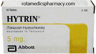
Cheap 5 mg hytrin fast delivery
Fong Y blood pressure medication usa buy cheap hytrin 2 mg on line, et al: Evidence-based gallbladder most cancers staging: changing cancer staging by analysis of information from the National Cancer Database arteria infraorbitalis hytrin 5 mg order amex, Ann Surg 243(6):767�771, dialogue 771-764, 2006. Franquet T, et al: Primary gallbladder carcinoma: imaging findings in 50 sufferers with pathologic correlation, Gastrointest Radiol 16(2):143� 148, 1991. Frauenschuh D, et al: How to proceed in patients with carcinoma detected after laparoscopic cholecystectomy, Langenbecks Arch Surg 385(8):495�500, 2000. Harder J, et al: Outpatient chemotherapy with gemcitabine and oxaliplatin in patients with biliary tract most cancers, Br J Cancer 95(7):848� 852, 2006. Hyder O, et al: Impact of adjuvant exterior beam radiotherapy on survival in surgically resected gallbladder adenocarcinoma: a propensity score-matched Surveillance, Epidemiology, and End Results analysis, Surgery 155(1):85�93, 2014. Imazu H, et al: Contrast-enhanced harmonic endoscopic ultrasonography within the differential diagnosis of gallbladder wall thickening, Dig Dis Sci 59(8):1909�1916, 2014. Ito H, et al: Polypoid lesions of the gallbladder: diagnosis and follow-up, J Am Coll Surg 208(4):570�575, 2009. Itoi T, et al: Detection of telomerase activity in biopsy specimens for prognosis of biliary tract cancers, Gastrointest Endosc 52(3):380�386, 2000. Jain K, et al: Sequential occurrence of preneoplastic lesions and accumulation of loss of heterozygosity in sufferers with gallbladder stones counsel causal association with gallbladder most cancers, Ann Surg 260(6):1073�1080, 2014. Javle M, et al: Molecular characterization of gallbladder cancer using somatic mutation profiling, Hum Pathol 45(4):701�708, 2014. Kato S, et al: Septum formation of the frequent hepatic duct associated with an anomalous junction of the pancreaticobiliary ductal system and gallbladder cancer: report of a case, Surg Today 24(6):534�537, 1994. Kimura W, et al: Clinicopathologic research of asymptomatic gallbladder carcinoma discovered at autopsy, Cancer 64(1):98�103, 1989. Koda M, et al: Expression of Fhit, Mlh1, and P53 protein in human gallbladder carcinoma, Cancer Lett 199(2):131�138, 2003. Kondo S, et al: Extensive surgery for carcinoma of the gallbladder, Br J Surg 89(2):179�184, 2002. Kozuka S, et al: Relation of adenoma to carcinoma in the gallbladder, Cancer 50(10):2226�2234, 1982. Kubota K, et al: How should polypoid lesions of the gallbladder be handled in the era of laparoscopic cholecystectomy Kumar S, et al: Infection as a risk factor for gallbladder most cancers, J Surg Oncol 93(8):633�639, 2006. Lee J, et al: Gemcitabine and oxaliplatin with or without erlotinib in superior biliary-tract cancer: a multicentre, open-label, randomised, Phase three study, Lancet Oncol 13(2):181�188, 2012. Matsumoto Y, et al: Surgical remedy of primary carcinoma of the gallbladder based on the histologic evaluation of 48 surgical specimens, Am J Surg 163(2):239�245, 1992. Naito Y, et al: Usefulness of lavage cytology throughout endoscopic transpapillary catheterization into the gallbladder within the cytological prognosis of gallbladder illness, Diagn Cytopathol 37(6):402�406, 2009. Naitoh I, et al: Unilateral versus bilateral endoscopic metallic stenting for malignant hilar biliary obstruction, J Gastroenterol Hepatol 24(4):552� 557, 2009. Nakamura S, et al: Aggressive surgery for carcinoma of the gallbladder, Surgery 106(3):467�473, 1989. Nakayama F: Recent progress within the analysis and treatment of carcinoma of the gallbladder: introduction, World J Surg 15(3):313�314, 1991. Nakazawa K, et al: Amplification and overexpression of c-erbB-2, epidermal development issue receptor, and c-met in biliary tract cancers, J Pathol 206(3):356�365, 2005. Ogura Y, et al: Radical operations for carcinoma of the gallbladder: current standing in Japan, World J Surg 15(3):337�343, 1991. Onoyama H, et al: Extended cholecystectomy for carcinoma of the gallbladder, World J Surg 19(5):758�763, 1995. Ouchi K, et al: Laparoscopic cholecystectomy for gallbladder carcinoma: outcomes of a Japanese survey of 498 patients, J Hepatobiliary Pancreat Surg 9(2):256�260, 2002. Pandey M: Environmental pollutants in gallbladder carcinogenesis, J Surg Oncol 93(8):640�643, 2006. Pandey M, et al: Carcinoma of the gallbladder: function of sonography in prognosis and staging, J Clin Ultrasound 28(5):227�232, 2000. Paolucci V, et al: Tumor seeding following laparoscopy: worldwide survey, World J Surg 23(10):989�995, discussion 996-997, 1999. Petrowsky H, et al: Impact of integrated positron emission tomography and computed tomography on staging and administration of gallbladder cancer and cholangiocarcinoma, J Hepatol 45(1):43�50, 2006. Principe A, et al: Radical surgery for gallbladder carcinoma: prospects of survival, Hepatogastroenterology 53(71):660�664, 2006. Rajagopalan V, et al: Gallbladder and biliary tract carcinoma: a comprehensive update. Randi G, et al: Gallbladder cancer worldwide: geographical distribution and risk factors, Int J Cancer 118(7):1591�1602, 2006. Rashid A: Cellular and molecular biology of biliary tract cancers, Surg Oncol Clin N Am 11(4):995�1009, 2002. Razumilava N, et al: Cancer surveillance in patients with major sclerosing cholangitis, Hepatology 54(5):1842�1852, 2011. Roa I, et al: Preneoplastic lesions and gallbladder most cancers: an estimate of the interval required for progression, Gastroenterology 111(1):232� 236, 1996. Roa I, et al: Gallstones and gallbladder cancer-volume and weight of gallstones are associated with gallbladder cancer: a case-control study, J Surg Oncol 93(8):624�628, 2006. Rodriguez-Fernandez A, et al: Application of contemporary imaging strategies in analysis of gallbladder cancer, J Surg Oncol 93(8):650�664, 2006. Sakamoto Y, et al: Clinical significance of extrahepatic bile duct resection for superior gallbladder cancer, J Surg Oncol 94(4):298�306, 2006. Sasaki R, et al: Hepatopancreatoduodenectomy with extensive lymph node dissection for locally advanced carcinoma of the gallbladder�longterm outcomes, Hepatogastroenterology 49(46):912�915, 2002. Sasatomi E, et al: Precancerous conditions of gallbladder carcinoma: overview of histopathologic traits and molecular genetic findings, J Hepatobiliary Pancreat Surg 7(6):556�567, 2000. Sato M, et al: Localized gallbladder carcinoma: sonographic findings, Abdom Imaging 26(6):619�622, 2001. Serra I, et al: Risk elements for gallbladder cancer: a global collaborative case-control study, Cancer 78(7):1515�1517, 1996. Sharma A, et al: Best supportive care compared with chemotherapy for unresectable gall bladder most cancers: a randomized managed research, J Clin Oncol 28(30):4581�4586, 2010a. Shimizu Y, et al: Should the extrahepatic bile duct be resected for regionally advanced gallbladder cancer Shindoh J, et al: Tumor location is a strong predictor of tumor progression and survival in T2 gallbladder cancer: an international multicenter research, Ann Surg 2014. Shinkai H, et al: Surgical indications for small polypoid lesions of the gallbladder, Am J Surg 175(2):114�117, 1998. Shirai Y, et al: Inapparent carcinoma of the gallbladder: an appraisal of a radical second operation after easy cholecystectomy, Ann Surg 215(4):326�331, 1992a. Shirai Y, et al: Radical surgery for gallbladder carcinoma: long-term results, Ann Surg 216(5):565�568, 1992b.
2 mg hytrin discount
Taki Y blood pressure medication kidney stones hytrin 2 mg purchase, et al: Predictive factors for enchancment of ascites after transjugular intrahepatic portosystemic shunt in sufferers with refractory ascites heart attack jaw pain right side buy hytrin 1 mg without a prescription, Hepatol Res 44(8):871�877, 2014. Tripathi D, et al: Good scientific outcomes following transjugular intrahepatic portosystemic stent-shunts in Budd-Chiari syndrome, Aliment Pharmacol Ther 39(8):864�872, 2014. Tripathi D, Jalan R: Transjugular intrahepatic portosystemic stentshunt in the management of gastric and ectopic varices, Eur J Gastroenterol Hepatol 18(11):1155�1160, 2006. Tsauo J, et al: Three-dimensional path planning software-assisted transjugular intrahepatic portosystemic shunt: a technical modification, Cardiovasc Intervent Radiol 38(3):742�746, 2015. Hepatic Cirrhosis, Portal Hypertension, and Hepatic Failure Chapter 87 Transjugular intrahepatic portosystemic shunting: indications and technique1247. Vidal V, et al: Usefulness of transjugular intrahepatic portosystemic shunt in the management of bleeding ectopic varices in cirrhotic patients, Cardiovasc Intervent Radiol 29(2):216�219, 2006. Wils A, et al: Transjugular intrahepatic portosystemic shunt in sufferers with persistent portal vein occlusion and cavernous transformation, J Clin Gastroenterol 43(10):982�984, 2009. Wu X, et al: Clinical outcome utilizing the fluency stent graft for transjugular intrahepatic portosystemic shunt in patients with portal hypertension, Am Surg 79(3):305�312, 2013. Xue H, et al: Transjugular intrahepatic portosystemic shunt vs endoscopic therapy in preventing variceal rebleeding, World J Gastroenterol 18(48):7341�7347, 2012. Yang Z, et al: Patency and medical outcomes of transjugular intrahepatic portosystemic shunt with polytetrafluoroethylene-covered stents versus bare stents: a meta-analysis, J Gastroenterol Hepatol 25(11): 1718�1725, 2010. Zhang F, et al: Different scoring methods in predicting survival in Chinese sufferers with liver cirrhosis undergoing transjugular intrahepatic portosystemic shunt, Eur J Gastroenterol Hepatol 26(8):853� 860, 2014. Orloff Among the etiologies of portal hypertension, these brought on by postsinusoidal obstruction are seen occasionally by most clinicians. Nonetheless, these disease processes represent complicated scientific challenges and require an intensive knowledge of the out there diagnostic and treatment modalities. The latter situation can be referred to as sinusoidal obstruction syndrome and is most frequently seen after myeloablation with chemotherapy or radiation therapy before hematopoetic stem cell transplant. For sufferers in whom these measures fail, liver transplantation stays a viable choice, with wonderful results despite recurrent illness in some reviews. Many facilities have adopted a stepwise strategy to therapy that has converted this once uniformly fatal process to a well-controlled, manageable condition. In reality, many experts agree that there may be multiple predisposing risk factors for the event of this syndrome. Although a short dialogue of this scientific phenomenon first appeared in a e-book by Budd in 1845, Lambron in 1842 is alleged to have reported the first case. In 1899, Chiari collected 10 circumstances and reported three private instances and presented the primary thorough clinicopathologic description of the syndrome, together with the speculation that the underlying mechanism was endophlebitis of the hepatic veins. The weight of evidence, however, favors the current opinion that the first course of is often thrombotic rather than inflammatory. Obstruction of hepatic venous outflow produces intense congestion of the liver and the scientific manifestations of ascites, hepatomegaly, and belly pain. In Western countries, a rapid course is common, and the outcome is commonly deadly in many reported cases. With immediate analysis and improved therapeutics, however, this situation can be managed as a chronic situation or cured entirely. Effective surgical remedy developed at extremely specialised facilities allows sturdy decompression of the obstructed hepatic vascular mattress. A review of reported instances signifies myelodysplasia as an underlying etiology in roughly half of affected patients (DeLeve et al, 2009). First, it was discovered more usually in younger adults, somewhat than in middle-aged and elderly sufferers. Second, polycythemia vera has been proven to be aware of treatment with hydroxyurea, which should be began as soon as the disease is discovered and continued for life. Whatever remedy regimen is used, the disease runs a benign course if treated early. The evaluation is an enlargement of the workup proposed by Mahmoud and Elias (1996) and others (Hirschberg et al, 2000; Valla, 2009) (Box 88. Pregnancy and Postpartum Budd-Chiari syndrome has been observed in women during being pregnant and, more typically, in the course of the postpartum interval. Axial (A) and coronal (B) magnetic resonance photographs reveal leiomyosarcoma (white arrows) of the intrahepatic inferior vena cava (white arrowhead) with nonopacification of the obstructed right hepatic vein. A congenital explanation for this condition has been proposed, however proof strongly suggests it represents the tip result of acquired thrombosis (Kage et al, 1992; Okuda, 2002; Okuda et al, 1995). Pathology the liver receives roughly one-fourth of the cardiac output via its twin afferent blood provide: the portal vein and hepatic artery. Obstruction to the egress of blood from the liver at any level alongside the outflow route ends in numerous severe hemodynamic and morphologic alterations. There is a marked improve in intrahepatic strain, which is reflected by an analogous improve in portal strain (see Chapter 76). The increased intrahepatic strain causes extravasation of plasma from the liver sinusoids and lymphatics with formation of ascites (see Chapter 81). Liver biopsy demonstrating typical traits of venous outflow obstruction under low (A) and excessive (B) energy. Note the marked sinusoidal congestion (black arrowhead) and hepatocyte atrophy (black arrow). With persistence of the obstruction, the necrotic parenchyma is changed by fibrous tissue and regenerating nodules of liver tissue. This pathophysiology is just like that seen after liver transplantation within the affected person with anastomotic venous outflow obstruction, with similar scientific manifestations. Early in the center of the illness, reduction of the obstruction may be expected to lead to reversal of the parenchymal and hemodynamic abnormalities. Late in the course, the damage to the hepatic parenchyma becomes irreversible; thus the timing of remedy has profound implications for the prognosis. The thrombus undergoes group and in the end is transformed to fibrous tissue that completely occludes the veins. Although recanalization of the occluded veins generally occurs, it hardly ever ends in efficient new outflow channels. Indeed, continual congestion of the liver leads to some extent of irreversible parenchymal harm. Retrograde propagation of the thrombus into smaller hepatic veins is usually discovered. Most of the circumstances have run a persistent course before discovery, and when first seen by a doctor, patients have in depth hepatic fibrosis or cirrhosis with portal hypertension and all its manifestations. Patients can have an acute or subacute course (typical for sufferers in Western countries), with fast progression of liver illness and its penalties during a number of weeks to a few months.
Proven 1 mg hytrin
It is thus not surprising that the pancreatoenteric anastomosis has intrigued surgeons blood pressure medication pros and cons order 5 mg hytrin overnight delivery, motivating them to seek for a extra reliable technique to avoid this dreaded complication blood pressure entry chart discount hytrin 1 mg otc. Many techniques have been described, and the literature will proceed to report novel methods that promise to be even safer. For so lengthy as the basic tenets of a secure anastomosis are met-namely, careful dealing with of the pancreatic tissues, a tension-free adaptation, good perfusion, and no distal obstruction-any pancreatoenteric anastomotic technique can have a great outcome. One of probably the most generally used techniques is a pancreaticojejunal anastomosis, which could be carried out by invaginating the transected pancreas into the tip of the jejunum, the so-called dunking procedure. Another variant is to anastomose the pancreatic duct on to a proper opening within the jejunum, the so-called duct-to-mucosa approach. The technique of pancreaticojejunal anastomosis-whether end-to-side or end-to-end, duct-to-mucosa or dunking-does not seem to significantly affect the anastomotic leak price. This can be accomplished by way of the ligament of Treitz or via a defect in the mesocolon just to the proper of the center colic vessels. The authors favor the latter because the jejunal limb is theoretically less weak to obstruction in the face of an area recurrence. The pancreatic remnant must be mobilized off the splenic vein for roughly 2 cm. The beforehand positioned hemostatic sutures previous to neck transection are used to barely elevate the gland away from the venous confluence. The first layer consists of transpancreatic horizontal mattress sutures positioned through and through the pancreas roughly 2 cm proximal to the minimize transection edge. It could also be helpful to straighten the needle to enable for a perpendicular path of the suture via the gland. It is important to begin 2 cm away from the edge anteriorly and likewise to exit the gland posteriorly the identical distance away from the transection edge. Next, a small seromuscular horizontal mattress suture is positioned via the jejunal limb close to the mesentery roughly 3 cm away from the stapled transected fringe of the bowel. The suture is then handed once more via the pancreas, coming into the posterior surface and exiting the anterior surface, once more maintaining 2 cm away from the minimize edge. In most circumstances, the pancreas will accommodate three such stitches inferior to the pancreatic duct and three superior. A probe must be placed in the pancreatic duct to defend it from being occluded whereas putting the horizontal mattress sutures. The needles should be left on every suture, as this stitch will be used for the ultimate anterior layer to full the anastomosis. Once this initial layer is complete, consideration is turned towards the duct-to-mucosa inside layer. The enterotomy must be placed roughly 1 cm away from the road of horizontal sutures to permit a tension-free duct-to-mucosa adaptation. Normally, four to eight sutures are capable of be placed to totally approximate the duct to jejunum, with two at the corners and one to three positioned anteriorly and posteriorly. Once the posterior layer is full and tied down, the corner sutures and anterior row can be positioned by way of the jejunum to full the anterior layer. For essentially the most inferior sew, the suture is placed in a vertical trend via the jejunum about 1 cm inferior to the pancreatic edge, guiding the needle anteriorly. These horizontal mattress sutures are continued, working from the inferior edge to probably the most superior sew. The corner and anterior row stitches positioned in the pancreatic duct to tent the duct open to facilitate placement of the posteriorrow. Endocrine Tumors Chapter sixty six Techniques of pancreatic resection 1017 fashion and then a horizontal mattress stitch directed again toward the pancreas. These vertical stitches serve to wrap the jejunum across the inferior and superior fringe of the pancreas. The end-to-side technique allows the variation of the jejunal opening to the precise requirements of the pancreatic remnant. Separate duct-to-mucosa adaptation also keeps the duct orifice open, thereby ensuring the unobstructed flow of pancreatic secretions by way of the anastomosis. Bilioenteric Anastomosis the bilioenteric anastomosis has less variability in its construction. It may be customary with a steady or interrupted approach, relying on the scale of the bile duct (see Chapter 31). For a steady approach, a singlelayer full-thickness anastomosis is constructed. A second suture is then used to full the anterior layer to forestall a pursestringing of the anastomosis. If an interrupted technique is chosen, which is the preferred approach for small bile ducts lower than 5 mm in diameter, the anterior row and nook sutures are first placed within the bile duct. This serves to maintain the duct open to facilitate development of the posterior row, similar to the discussion for the pancreaticojejunostomy. A small enterotomy is made in the jejunum, approximately 6 to 8 cm away from the pancreaticojejunostomy. The posterior bile duct sutures are then placed and handed by way of full-thickness jejunum as properly. The anterior row and nook sutures are then handed through full-thickness jejunum. Once this anastomosis is complete, the jejunum is tacked to the mesocolon to relieve rigidity off the reconstruction and prevent herniation of the small bowel into the proper higher quadrant. One group speculated that a retrocolic reconstruction predisposes the jejunal limb to venous congestion and bowel edema, which may consequently retard recovery of jejunal peristalsis at the duodenojejunostomy (Park et al, 2003). Postoperative gastroparesis can also result in momentary gastric distension, which may result in angulation of the anastomosis because it lies comparatively fixed via its retrocolic place (Horstmann et al, 2004). None of the duct-to-mucosa sutures should be seen as quickly as the anterior row is complete, and the jejunum is folded over the anterior surface of the pancreas. Interrupted suture technique for bilioenteric anastomosis exhibiting completed posterior row with nook and anterior bile ductsuturesinplace. The surgical procedure of choice for tumors arising in the physique or tail of the pancreas is a distal pancreatectomy. This operation entails the removing of that portion of the pancreas extending to the left of the midline, not including the duodenum and distal bile duct. Technique Following a thorough examination of the peritoneal cavity, the gastrocolic ligament is split sufficiently to allow full visualization of the pancreas to its tail and the hilum of the spleen. The point of division is chosen within the proximal normal pancreas, and the overlying peritoneum is split alongside the superior and inferior borders of the pancreas. The inferior border of the pancreas is isolated, with subsequent inferior mobilization of the splenic flexure. Once the pancreas has been uncovered and the brief gastric vessels have been ligated, consideration should be paid to the three buildings (splenic artery, splenic vein, and pancreatic parenchyma) that shall be divided. The actual technique for pancreatic transection and stump closure has been the subject of a lot debate.
5 mg hytrin order fast delivery
In summary arteria braquial 2 mg hytrin generic overnight delivery, liver metastases regularly present in synchronous blood pressure medication that does not lower heart rate discount 2 mg hytrin amex, multifocal, and bilateral configurations. The variety of displays of liver metastases, by method of size, number, and placement, demand that the liver surgeon not be limited to anyone method or method. With the probe aligned perpendicular to a cautery-scored capsular mark, the image demonstrates the space from the massive tumor (hyperechoic white area) and hepatic veins (central anechoic structures) to the proposed transection line (linear hypoechoic shadow). After main hepatectomy, interrogation of the safety of bile duct closure on the transection surface has been proven to cut back significantly the danger of clinically important postoperative bile leak (Zimmitti et al, 2013a). Not solely does synchronous metastatic illness portend a worse prognosis than metachronous metastases (Scheele et al, 1995), however liver and colorectal surgeons also are regularly pressed to make critical preliminary therapy choices without the good factor about knowing the response to systemic therapy. The second decision level is to decide the surgical therapy sequence: liver first, conventional colon first, or simultaneous resection. Several elements associated to perioperative and long-term outcome should be thought of when deciding among these options. With regard to surgical morbidity, multiple teams have reported a lower complication rate in patients treated with a single simultaneous surgical procedure compared to the aggregated morbidity of two separate resections (Capussotti et al, 2007; Chua et al, 2004; de Haas et al, 2010; Martin et al, 2003; Vogt et al, 1991). However, the vast majority of patients within the simultaneous-surgery arms of these research required only a minor hepatectomy. Patients with extra complex liver illness show a significant improve in major morbidity if liver resection is paired with or follows the "contaminated" colectomy process. Hypokalemia and hypophosphatemia happen inside forty eight hours, because the regenerating liver begins to consume these electrolytes. A, Portogram, and B, 1-month postprocedure computed tomographic scan, after portal vein embolization to facilitate resection of colorectal liver metastases. The fluid rate could be titrated to keep hourly urine output between 25 and 50 mL. The acute surgical trauma results in an early, high-spiking lactate dehydrogenase stage. Rapid return towards regular by postoperative day 2 indicators liver recovery and low probability for liver failure. In fact, posthepatectomy sufferers are at a lot larger danger of thrombotic events than bleeding occasions, and the danger is immediately proportional to the magnitude of hepatectomy (Tzeng et al, 2012). In challenging cases, thromboelastography should be used to guide blood product therapy. Subsequent architectural rearrangements continue over the following few weeks, and within the majority of circumstances, this process is complete in 6 weeks. During this phase of recovery, sufferers sometimes report fatigue and early satiety. Mortality the mortality related to an elective liver resection for colorectal metastases has decreased significantly over the previous 3 a long time, and in the last 10 years, it has been proven to be less than 3% throughout major series. Advances in the understanding of liver anatomy, resection strategies, and anesthetic care have translated into favorable mortality rates, even with major resections. The majority of posthepatectomy deaths happen from perioperative hemorrhage or liver failure. Morbidity the complication fee associated with hepatectomy is attributable in giant part to the metabolic and immunologic stress associated with liver surgery. Most series report that approximately 50% of patients will expertise a complication, and as many as 20% will expertise a major complication (Table 92. Extrahepatic morbidities embody cardiac, pulmonary, and infectious complications. The incidence of cardiovascular complications is about 9%, but the majority of those are arrhythmogenic in nature (Jarnagin, 2002). The low price of extreme cardiac problems displays careful choice of patients for liver resection. Pulmonary complications happen in virtually 20% of sufferers and are associated to the big higher stomach incisions, postsurgical sympathetic pleural effusions, and failure to mobilize sufferers early. Specifically, symptomatic pleural effusions occur in 10% of cases, pneumonia in 3%, and pulmonary embolism in 1% (Jarnagin, 2002). Liver insufficiency and liver failure remain the most dangerous liver-related issues and happen in 3% to 8% of sufferers with main hepatic resection (Balzan et al, 2005, Rahbari et al, 2011). Other hepatobiliary problems include bile leak in 4% of patients and perihepatic abscess in 2% to 10% (Jarnagin, 2002; Zimmitti et al, 2013a). Significant hemorrhage is uncommon (1%-3%) but can be an necessary cause of early perioperative mortality. To assess and compare postoperative issues precisely, morbidity, Perioperative Morbidity and Mortality For a surgical approach to a metastatic disease to be widely accepted, it have to be possible, secure, and effective. Over time, vital technical enhancements in surgery and anesthesia have allowed main liver resection to be carried out with acceptable morbidity and minimal mortality. These procedures, nevertheless, stay at the upper finish of the chance spectrum in general surgery. Using correct patient choice, preoperative staging, and surgical expertise, these dangers can be minimized, leading to extended disease-free survival and attainable remedy. For instance, in the expertise of Jarnagin (2002), with more than 1800 liver resections performed at a single heart, the median hospital keep was 8 days, and intensive care unit admission was required for less than 112 patients (6%). More lately, reported postoperative length-of-stay values have declined, with patients who obtain minimally invasive approaches or enhanced recovery protocols meeting discharge criteria in 2 to four days. Choti (1998) and Glasgow (1999) and colleagues used state registries in Maryland and California, respectively, and located that hepatectomy performed at a high-volume heart was associated with reductions in perioperative mortality, length of hospital keep, and value (Choti et al, 1998; Glasgow et al, 1999). A study by Fong and colleagues (2005), based mostly on a national Medicare database, offers further support for superior outcomes at high-volume centers. The authors demonstrated that perioperative outcomes correlated with the expertise of the institution, and a survival advantage endured after the perioperative interval. These knowledge lend assist to the concept of regionalization of hepatectomy to high-volume facilities. As adjuvant remedy has improved, patients with R1 resections frequently experience outcomes that more closely method sufferers with negative resection margins. With regard to the optimum or acceptable width of unfavorable margin, earlier research indicated that a 1-cm rim of normal liver parenchyma surrounding a resected tumor was ideal (Shirabe et al, 1997). Further supporting this concept, two Japanese microdissection research have demonstrated low charges of micrometastastic disease, or satellitosis, in resection specimens (Kokudo et al, 2002; Yamamoto et al, 1995). Impact of Postoperative Complications Higher operative blood loss and transfusion requirements have been associated with an elevated number of perioperative complications and mortality, however not with a worse long-term survival (Kooby et al, 2003b). On the opposite hand, an affiliation between the development of other postoperative issues and decreased long-term outcomes (both disease-free and overall survival) has been observed for liver surgical procedure (Farid et al, 2010; Ito et al, 2008). This finding is supported by a large, population-based evaluation of sufferers undergoing surgical procedures (Khuri et al, 2005), underscoring the importance of security in oncologic liver surgery.
Diseases
- Glucosidase acid-1,4-alpha deficiency
- Hirschsprung disease
- Gray platelet syndrome
- Leigh syndrome, French Canadian type
- Chromosome 4, partial trisomy distal 4q
- Grant syndrome
- Gougerot Sjogren syndrome
- Rosenberg Lohr syndrome
- Polycystic ovarian disease, familial
5 mg hytrin cheap with visa
Of these eighty two patients blood pressure eyes hytrin 2 mg amex, only six (7%) have been resectable arrhythmia jaw pain 2 mg hytrin buy otc, and all six patients skilled recurrence or died of illness inside 2 years. The median survival in jaundiced sufferers was 6 months in contrast with 16 months in patients presenting with out jaundice. Thus jaundice, consultant of regionally superior disease, may be a sign for therapy with chemotherapy earlier than consideration for resection. A report from New York Hospital reviewed their experience with gallbladder cancer from 1915 via 2000 (Grobmyer et al, 2004). Throughout the years, the presentation was remarkably comparable in that the majority patients had superior disease at presentation. Over time, nonetheless, a lower was seen in the proportion of sufferers who offered with weight reduction (42% vs. The percentage of sufferers presenting with stomach ache and jaundice was related, roughly 50% and 85%. Overall, serum tumor markers are of minimal medical worth in contrast with clinical awareness, a heightened degree of suspicion in acceptable circumstances, and good-quality imaging research. Because most patients current with superior illness, it is very important try to set up the diagnosis and the C. Malignant Tumors Chapter 49 Tumors of the gallbladder 793 extent of illness with imaging to reduce the variety of sufferers who should bear a nontherapeutic surgical exploration. Except for the earliest-stage illness, this should now be potential in most sufferers. It is rare, nevertheless, to find pulmonary metastases with out regionally advanced or intraabdominal metastatic disease (Lee et al, 2010). Ultrasonography is a superb imaging modality for the gallbladder (see Chapter 15). Findings such as discontinuous mucosa, echogenic mucosa, and submucosal echolucency are more widespread in early malignancy compared with benign gallbladder illness. Doppler assessment of blood move through areas of mucosal abnormalities may help to differentiate early malignancy from benign disease, and contrast-enhanced ultrasound strategies might improve detection confidence even additional (Choi et al, 2013; Imazu et al, 2014; Sato et al, 2001). In one research of gallbladder cancer patients, a polypoid mass was present in 27% of circumstances, and a gallbladder-replacing or invasive mass was current 50% of the time (Wibbenmeyer et al, 1995). Ultrasound is an effective modality for evaluating the direct extension of gallbladder most cancers. One retrospective research reported that in 203 patients with gallbladder cancer, a mass was identified in 177 sufferers (87%) on preoperative ultrasound (Pandey et al, 2000). Ultrasound was limited, nonetheless, in identifying lymph node metastases in pericholedochal and peripancreatic nodes. Because most cases are superior, the most common discovering in gallbladder cancer is a heterogeneous mass changing all or a half of the gallbladder (Bach et al, 1998; Franquet et al, 1991). Diffuse thickening of the gallbladder wall also is a standard finding on cross-sectional imaging and on ultrasound, but this can be difficult to differentiate from benign inflammatory modifications. These strategies present essential information about the native extent of disease and whether or not distant metastases are current. The overall accuracy improved from 72% to 85% when multiplanar reconstructions have been added to typical axial imaging. The false-positive case was in a affected person with xanthogranulomatous cholecystitis (Koh et al, 2003). Duplex ultrasound adds information when it comes to vascular tumors and helps assess native hepatic vasculature. Gallbladder cancer presenting with jaundice is relatively common, and this diagnosis must be in the differential analysis for any malignant-appearing, mid�bile duct stricture. One additionally should contemplate gallbladder most cancers when the prognosis of Mirizzi syndrome (see Chapter 42) has been made (Redaelli et al, 1997). Based on the extent of obstruction, a judgment may be made as to one of the best strategy for biliary stenting (endoscopic retrograde vs. As mentioned later, this can be a crucial part of the palliation of superior gallbladder most cancers. Gallbladder cancer has a tendency to seed the peritoneum, biopsy tracts, and surgical wounds (Fong et al, 1993; Hu et al, 2008; Merz et al, 1993), and pointless biopsies merely increase this risk. It is crucial that if the analysis is suspected, the surgeon and affected person must be ready for a definitive operation. The surgeon and patient also should be ready for the possibility of performing a liver resection for benign illness. In skilled hands, a limited liver resection ought to be secure, and the chance of this process must be lower than the risk of a quantity of biopsies or noncurative operations. For a patient with unresectable or metastatic illness, a percutaneous biopsy has an accuracy of just about 90%, and the falsepositive rate is negligible (Akosa et al, 1995). Bile cytology has been proposed as a way of constructing the prognosis of gallbladder cancer without risking peritoneal seeding. Sensitivity of bile cytology has been reported to be approximately 75% (Akosa et al, 1995; Arora et al, 2005; Mohandas et al, 1994; Naito et al, 2009). It utilizes tumor depth (T), nodal standing (N), and the presence of metastases (M) to place patients into four levels based on pathologic and radiographic findings. T1a tumors invade the lamina propria of the gallbladder; T1b tumors invade the muscular layers. T2 tumors unfold by way of the muscular layers into, however not past, the connective tissue layers. Anatomically, this interprets to unfold into the gallbladder serosa or cystic plate, however not the liver parenchyma. T3 lesions spread via the gallbladder serosa and invade the liver, extrahepatic bile ducts, or perihepatic organs. T4 lesions invade multiple extrahepatic organs and/or major vessels and generally reflect domestically unresectable disease. N1 nodes are lymph nodes adjoining to the cystic duct, bile duct, hepatic artery, and portal vein. The only polypoid lesions that have malignant potential and are associated with a significant price of harboring malignancy are adenomatous polyps. Adenomyomatosis, outlined as extension of Rokitansky-Aschoff sinuses via the muscular wall, is widespread and often diagnosed by ultrasound criteria (Stunell et al, 2008). The relevant clinical query is which lesions mandate a cholecystectomy in an asymptomatic affected person. Numerous scientific reviews have identified elements associated with malignancy in gallbladder polyps. The most consistent predictors are single polyps, size larger than 1 cm, and age older than 50 (Shinkai et al, 1998; Yeh et al, 2001). Although some authors have really helpful cholecystectomy for any patient with fewer than three polyps (Shinkai et al, 1998), we usually advocate cholecystectomy for any polyp larger than 1 cm as a outcome of the chance of malignancy for polyps lower than this dimension, regardless of number, is exceedingly low. Eighty sufferers underwent cholecystectomy, and a single patient had carcinoma in situ in a 13 mm gallbladder polyp. The exception to this recommendation of resection only for polyps larger than 1 cm is for these arising within the setting of major sclerosing cholangitis. The prevalence of gallbladder most cancers is higher in this affected person population, and polyps past 0.
5 mg hytrin with amex
Lymph node metastases and extrahepatic extension had been additionally related to poorer outcome hypertension lowering foods hytrin 1 mg amex. Two current publications also emphasised the importance of tumor measurement and differentiation (Ali et al blood pressure effects purchase 5 mg hytrin with visa, 2015; Spolverato et al, 2014). Resection of up to 80% of hepatic quantity may be contemplated in sufferers with good liver perform and as much as 60% in patients with compromised liver perform (Ebata et al, 2012). Often, nevertheless, resections of this magnitude might need to be preceded by portal vein embolization (Shindoh et al, 2013). Resection with positive margins or residual macroscopic disease is related to median survivals of 1. In contrast, 5-year survival rates after full resection vary between 13% and 43% (Table 50. The principal reason for the variability in survival seems to be the presence of lymph node metastases. Lieser and colleagues (1998) reported 5-year survival of 42%, however solely 13% of patients presented with lymph node metastases, whereas within the series of Chu and Fan (1999), 50% of sufferers introduced with nodal metastases, and none survived 5 years. All these medical collection emphasize the prognostic importance of acquiring an R0 resection, which often requires a serious hepatic resection. Cherqui and colleagues (1995) achieved related results with an aggressive surgical policy and liberal use of major prolonged hepatectomy. Status of Lymphadenectomy Although the significance of reaching an R0 resection is clear, the function of routine lymph node dissection is debated. The presence and extent of nodal metastatic illness are necessary prognostic factors. Chu and Fan (1999) dissected portal lymph nodes in their series, and all patients with portal lymph node metastases died within 10 months of resection. A additional examine from Japan (Shirabe et al, 2002) confirmed that there was no survival benefit for patients undergoing hepatectomy with portal lymphadenectomy versus hepatectomy alone. However, Nozaki and colleagues (1998) recommended routine dissection of cardia and lesser curvature nodes for left-sided tumors and dissection of the hepaticoduodenal ligament for right-sided tumors, though using this strategy was not related to variations in survival. Extended surgical procedure has been related to higher mortality (Yamamoto et al, 1999). Despite these findings, nevertheless, latest series present that greater than half of sufferers bear routine lymphadenectomy (De Jong et al, 2011; Ribero et al, 2012; Uchiyama et al, 2011). A systematic evaluation confirms a development toward routine lymph node dissection, with greater than 75% of sufferers present process lymphadenectomy (Aimini et al, 2014). In these series, the speed of lymph node positivity was between 30% (De Jong et al, 2011) and 45% (Aimini et al, 2014). In addition, Nakayama (2014) and Choi (2009) and colleagues instructed an related extended survival for node-positive patients present process lymphadenectomy. From Liver Cancer Study Group of Japan, 2003: General Rules for the Clinical and Pathological Study of Primary Liver Cancer, 2nd ed. The roles of neoadjuvant and adjuvant chemotherapy, each systemic and regional; conformal radiation therapy; and ablative therapies are beneath investigation (Weber et al, 2015). With respect to staging, uniform agreement is missing on the optimum variety of nodes harvested per affected person. Usually, three or much less are harvested (De Jong et al, 2011), although as a lot as seven nodes are instructed for patients with hilar cholangiocarcinoma (Ito et al, 2010). Thorough evaluation of all intraabdominal nodal basins must be undertaken before hepatic resection, and sampling of suspicious nodes is indicated to stage illness precisely, which may direct postoperative remedy. Most surgical collection affirm that the presence of lymph node metastases is the most important prognostic factor. Endo and colleagues (2008) documented a recurrence rate of 93% in node-positive patients present process R0 resection versus 47% in node-negative patients. These investigators also found that tumor measurement higher than 5 cm in diameter and the presence of multiple intrahepatic tumors have been significant adverse prognostic components. Other investigators have also defined lymphatic permeation, vascular invasion, and intrahepatic satellite tv for pc lesions as predictors of poor survival. After resection, the most common website of recurrence is within the liver, followed by intraabdominal tumor, pulmonary metastases, and bony metastases (Jan et al, 2005). These investigators reported a median survival of 5 months in 18 sufferers treated with liver transplantation, with a 1-year survival fee of thirteen. The patients in each stories have been deemed irresectable however had no evidence of extrahepatic unfold, and the authors concluded that transplantation could be thought-about on this group as a outcome of the results achieved are higher than palliative C. This is in distinction to the emerging protocol of neoadjuvant chemoradiation earlier than transplantation for hilar cholangiocarcinoma (Schwartz et al, 2009). Tumor Ablation Tumor ablation refers to the intrahepatic destruction of tumors using thermal energy. Historically, cryotherapy (see Chapter 98D) had been employed (Cuschieri et al, 1995; Sheen et al, 2002), however presently, radiofrequency (see Chapter 98B) and microwave (see Chapter 98C) are most often used. Since most of the lesions are giant at presentation, their size usually precludes efficient ablation. Likewise, the use of ablation to treat intrahepatic metastases is unwise as a result of these are markers of vascular invasion and diffuse illness. However, Rai and colleagues (2005) reported a case of recurrent tumor after transplantation treated with radiofrequency ablation and managed for 12 months. Also, a quantity of small collection (Kim et al, 2011; Xu et al 2012; Yu et al, 2011) have shown that complete tumor ablation can be achieved using percutaneous ablation in sufferers not appropriate for resection. In basic, small (<3 cm in diameter) solitary tumors are most fitted for this method, somewhat than multiple tumors or recurrent illness. In rigorously chosen sufferers, a 2-year survival rate of 60% has been reported in those that would otherwise be managed with best supportive care (Yu et al, 2011). Subsequently, Mouli and associates (2013) prospectively treated 46 sufferers, with a 98% response fee, median survival of 15 months, and five patients downstaged to turn into eligible for resection or transplant. Overall, 87% of sufferers achieved a response or had stable illness, with median survival of thirteen months. These outcomes were confirmed by a later study utilizing the same routine (Kiefer et al, 2011). Vogl and colleagues (2012) reported a big sequence of one hundred fifteen sufferers treated with mitomycin C alone, gemcitabine alone, mitomycin C and gemcitabine, or mitomycin C and cisplatin, with a complete of 819 chemoembolizations and 7 interventions per patient. These investigators obtained one hundred pc response fee in 11 sufferers, with median survival of 13 months. This reflects that cholangiocytes are multifunctional proliferative cells, produce stimulatory cytokines and act through autocrine and paracrine pathways, and in addition play a task in mediating irritation, a key issue in the initiation and upkeep of carcinogenesis. In addition, cholangiocytes even have a task in cleansing and excretion of metabolites into bile, which contributes to chemotherapy resistance. Thirty patients had neoadjuvant therapy, which significantly delayed surgical resection by a mean of 6. However, the randomized trial of Valle and colleagues (2010) established that a mix of gemcitabine and cisplatin was superior to gemcitabine alone for the treatment of biliary tract cancers in patients with domestically advanced and metastatic disease. Importantly, only 17% of sufferers on this series had R0 resection (Bhudhisawasdi et al, 2012).
Hytrin 1 mg buy cheap on line
Ensuing symptoms are often related to the migration of the mucinous cystic content material arteria ileocolica cheap 1 mg hytrin amex. Other problems have included rupture in the peritoneal cavity blood pressure 160100 purchase 1 mg hytrin fast delivery, superinfection, bleeding, and caval compression, as beforehand talked about. Management Cystadenomas require complete excision to stop recurrence of symptoms and malignant transformation. Evidence that partial excision, aspiration, and external or inner drainage are ineffective is that recurrence has been noted very early on (Devaney et al, 1994; Ishak et al, 1977; Lewis et al, 1988; Wheeler & Edmondson, 1985) and that 40% to 50% of the sufferers culled by tertiary referral centers had undergone such earlier treatments previous to referral (Daniels et al, 2006; Delis et al, 2008; Hansman et al, 2001; Thomas et al, 2005; Vogt et al, 2005). Recurrence occurs at a mean of 21 months but may be delayed as much as 4 years (Ahanatha Pillai et al, 2012; Vogt et al, 2005). This might clarify why recurrence following incomplete resection has not been systematically noticed, as most studies only have short-term follow-up (Barabino et al, 2004; Lewis et al, 1988; Manouras et al, 2008). Development of a cystadenoma following partial resection of a cystadenocarcinoma has additionally been documented (Akwari et al, 1990; Devine et al, 1985; Lei & Howard, 1992; Woods, 1981). Fenestration of the cystadenoma with fulguration of the interior cystic lining has been attempted with occasionally enough long-term success (Thomas et al, 2005), but experience is too restricted to suggest this technique. Treatment of extrahepatic cystadenoma ought to embrace bile duct resection and bilioenteric reconstruction quite than easy enucleation from the bile duct wall. As for different tumors, laparoscopic resection can be an option for trained surgeons (Koffron et al, 2004; Veroux et al, 2005). A single case of recurrence following full resection has been reported (Wheeler & Edmondson, 1985). This discordance is puzzling but probably speaks to the dearth of strict pathologic definition of this entity. The epithelium is phenotypically much like the classic cystadenoma (see earlier). Otherwise, age (peaking between 40 and 60 years), symptoms, and measurement of the cyst are comparable (Buetow et al, 1995). Percentage refers to the proportion of cystadenocarcinoma among cystadenomatous tumors. Case reports counsel that it may bear morphologic modifications with a progressive improve in diameter, mural thickening, growth of papillary nodules, and subsequent malignant transformation (Akiyoshi et al, 2003; Fukunaga et al, 2008). Single case reviews exist in which malignancy was an adenosquamous carcinoma (Devaney et al, 1994), or it developed in the stroma (Akwari et al, 1990). Cystadenocarcinoma was first described in 1943 by Willis, but by 2000, solely a hundred had been reported in the literature (Bardin et al, 2004; Lauffer et al, 1998), and they had been considered to account for under zero. Most have been discovered within the liver, with only anecdotal reviews of locations in the extrahepatic bile duct and the gallbladder specifically (Waldmann et al, 2006). This should be taken under consideration when analyzing the literature, and even when studying this part. Although often nonspecific, signs are nearly always current and are initially rather indolent (Xu et al, 2015). They are common to cystadenoma and embody stomach swelling, discomfort, ache, or palpation of an abdominal mass. Biliary obstruction with jaundice is reported in 20% of the sufferers, associated to biliary compression or migration of mucus or tumor materials. Acute symptoms may in any other case happen because of intracystic hemorrhage or tumor rupture. As a rule, prognosis is delayed, and the mean time interval between first signs and remedy is 29 months (Lauffer et al, 1998); one case was reported of a cystadenocarcinoma being found 1 yr after bone metastasis (Berjian et al, 1981). Origin and the CystadenomaCystadenocarcinoma Sequence Cystadenocarcinoma has no recognized risk factors, and its origin is unknown but is normally assumed to symbolize the malignant counterpart of cystadenoma. The proliferating epithelial cells certainly resemble those noticed in cystadenoma; each categorical a biliary-type phenotype, and typical areas of cystadenoma (benign columnar epithelium) coexist with malignant papillary epithelium in most sufferers (up to 90%; Lauffer et al, 1998). Longitudinal morphologic research of single patients whose cystic lesions had been adopted for more than a decade have additionally proven the progressive growth of typical cystadenocarcinoma (Akiyoshi et al, 2003; Kubota et al, 2003). As a whole, cystadenocarcinomas also are likely to be larger and to be discovered 5 to 10 years later than cystadenoma (Lauffer et al, 1998). The transition from one to the other is assumed to happen via dysplasia or metaplasia that may be noticed inside cystadenoma (see "Pathology" earlier). The risk of malignant transformation of cystadenoma into cystadenocarcinoma is unknown, as the tumor is uncommon, and restricted knowledge can be found. A tough estimate may be made by comparing the relative proportion of "cystadenoma" and cystadenocarcinoma in published sequence (see Table 90B. Although biased, this estimate is consistent with that reported for choledochal cysts, also premalignant lesions. Case reviews have proven that a lesion might remain morphologically unchanged for years, however that once mural nodules appear, these could progress within a couple of months (Akiyoshi et al, 2003). The propensity of, and time essential for, the malignant epithelial proliferation to turn out to be invasive can be unknown. It has been reported that in approximately one third (Devaney et al, 1994) to one half (Lauffer et al, 1998; Nakajima et al, 1992) of the patients who had undergone surgical procedure, the tumor was confined to the cyst, which may clarify the very excessive survival rates reported after full resection. However, parenchymal extension appearing as satellite tv for pc nodules or perineural and lymphatic invasion for tumors located near a glissonian pedicle does exist, and distant metastases are present on the time of analysis in 20% of the sufferers (Lauffer et al, 1998). Gross Morphology and Imaging Cystadenocarcinoma shares a lot of the morphologic and radiologic options of cystadenoma. The lesions are solitary (Seo et al, 2010) and usually massive, with a median diameter of 12 cm however rising as a lot as forty cm (Lauffer et al, 1998); they may also be small, and cystadenocarcinoma of lower than 5 cm have been reported, including one being 2 cm (Lauffer et al, 1998; Lewin et al, 2006; Williams et al, 2001). Macroscopically, they more regularly have a hemorrhagic or bilious content (Buetow et al, 1995), and hemorrhage or nodularity within the cyst wall is clear (Buetow et al, 1995; Lewin et al, 2006; Seo et al, 2010). However, all of these options can be noticed in nonmalignant cystadenoma (Choi et al, 2010; Fukunaga et al, 2008), and differentiating both tumors on macroscopy or imaging alone has as much as now confirmed very tough (Buetow et al, 1995; Hai et al, 2003). Diagnosis Cystadenocarcinoma could theoretically be mistaken for metastases from cystadenocarcinoma of the ovary or pancreas, however these are normally multiple, whereas cystadenocarcinomas are, as a rule, single lesions. It could be argued that these distinctions are of little sensible importance as all require resection; nonetheless, the extent of resection could differ. Cyst sampling should in any case be performed with caution, as cystadenocarcinomas, and biliary tumors generally, have a excessive propensity for peritoneal seeding that has been observed following this process (Iemoto et al, 1981; Nakajima et al, 1992). Pleural effusion, presumed to be associated to aspiration cytology, has also been reported (Hai et al, 2003). Resection of the biliary confluence adopted by a hepaticojejunostomy could additionally be required if the tumor lies near the biliary confluence. Every effort ought to be made not to open the cyst as peritoneal carcinomatosis has been noticed after unintended cyst rupture despite unfavorable cytology for tumor cells in cystic fluid (Kosuge et al, 1992). Following complete resection, the prognosis appears higher overall than that of patients with hepatocellular carcinoma (see Chapter 91) or cholangiocarcinoma (see Chapter 50). Following complete resection, 5-year survival may be as high as 65% to 70% (Lauffer et al, 1998; Thomas et al, 2005; Vogt et al, 2005; Xu et al, 2015). The ductectatic sort evidences diffusely dilated bile ducts, and the cystic sort seems as a large cystic mass (Sakamoto et al, 1997).
1 mg hytrin buy fast delivery
Some suggest that survival is best when both sides of the liver are drained (Chang et al heart attack zippy demi generic hytrin 1 mg mastercard, 1998) blood pressure rates chart 2 mg hytrin purchase with visa. In either case, we favor to stent into the frequent bile duct and not into the duodenum if possible so as to preserve operate of the sphincter of Oddi. One advantage of the extra anatomic Y-shaped configuration stent placement is that if stent occlusion occurs, each stents are approachable endoscopically. In this case, and within the absence of some other contraindication, a affected person being drained for pruritus alone might need a major stent positioned, as a result of only a small portion of the liver needs to be drained to alleviate pruritus. We use this as a rule of thumb, despite that in a series of 149 sufferers drained at Memorial Sloan Kettering Cancer Center, we found only a touch significant difference in the number of sufferers attaining a bilirubin lower than 2 mg/dL, based mostly on the estimated quantity of liver drained. In this analysis, 6 (29%) of 21 patients with less than one third of the liver drained attained a bilirubin level below 2 mg/dL, whereas this was achieved in sixty five (51%) of 128 sufferers with a couple of third of the liver drained (P =. After stent placement, if the bilirubin fails to fall to the desired level, a second drainage process can be performed. If an internal/external drainage catheter is placed, and subsequently the serum bilirubin normalizes with out evidence of cholangitis, the affected person can bear stenting of that portion of the liver drained by the catheter. When the initial drainage is on the right aspect, and the tumor has prolonged up the right hepatic duct in order to isolate the anterior and posterior divisions from each other and from the left hepatic duct, side-by-side self-expanding metallic stents could be placed on the proper to drain both the anterior and posterior ducts. Alternatively, when the left facet of the liver is practical, a left drainage may be performed as the next step; then one stent can be placed from the left, and another may be positioned from the best. Although a big difference in patency is reported when a couple of stent is placed in a noncoaxial method (Maybody et al, 2009), the mean patency of multiple stents is nearly 6 months, justifying stent placement. The ideas of biliary drainage are simple, however when excessive bile duct obstruction is current, the planning is complicated, and execution may be tough. The patient will need to have enough of the liver drained to be free of cholangitis and pruritus and to effect a discount in serum bilirubin to obtain chemotherapy, if indicated. Given that no distinction in stent patency is reported if the stent is inserted for proximal or distal obstruction, that a major distinction in patency is seen when a couple of stent is placed, and that lower complication charges are reported when stents are positioned primarily, major stent placement must be thought-about whenever possible (Inal et al, 2003a, 2003c; Maybody et al, 2009). With proper technique, together with peripheral bile duct puncture, serious bleeding problems are uncommon. Because the hepatic artery, portal vein, and bile duct journey facet by aspect inside portal triads, blood may enter the bile duct during catheter exchanges, leading to hemobilia in the immediate postprocedure interval (see Chapter 125). Hemobilia usually clears within 24 hours, and new or recurrent hemobilia within the first few days of drainage typically is said to catheter malposition. If the catheter is pulled out from its original place, a catheter sidehole might become positioned adjoining to a portal vein department; this problem could be corrected by simply repositioning the catheter, however the catheter is usually upsized as properly. No matter where the initial puncture is performed to opacify the biliary tree, makes an attempt are always made to puncture a peripheral bile duct for catheter placement, preferably a fourth-order or fifth-order branch. The more peripheral the bile duct punctured, the smaller the accompanying hepatic artery department, and the decrease the chance of arterial damage and postprocedure bleeding. Despite prophylactic antibiotic coverage, sepsis may occur immediately after or within a number of hours of drainage and should be handled appropriately (Smith et al, 2004). This is most frequently manifested by the development of rigors with normal or low body temperature, but hypotension and fever may also occur. Sepsis is managed with continued administration of appropriate antibiotics, expansion of intravascular volume, and pressor support if needed. Blood cultures must be drawn to establish organisms responsible for the bacteremia. This is particularly important for those with preprocedure fever, biliary-enteric anastomosis or sphincterotomy, earlier endoscopic retrograde cholangiopancreatography, or an indwelling stent or catheter. Although constructive bile cultures are more widespread in patients with benign bile duct obstruction, cultures are positive in more than half of patients with malignant obstruction. Five percent of sufferers without fever, earlier biliary surgical procedure, or endoscopic or percutaneous intervention have constructive bile cultures (Brody et al, 1998). Leaking is most frequently related to the catheter becoming malpositioned so that a number of sideholes are no longer inside the biliary tree however are within the catheter tract and even outdoors the affected person. Leakage may be seen with lack of adequate sideholes above the level of obstruction. Anything that impedes the circulate of bile from above the obstruction, both through the catheter to beneath the obstruction or right into a drainage bag, will lead to bile leaking again along a longtime tract. For a properly positioned catheter with an applicable number of sideholes, the issue is well remedied by catheter trade. Patients with capped internal-external catheters could have bile leak back along the catheter tract when egress of bile is obstructed internally. Distal sidehole occlusion is the most common trigger, and this problem is easily remedied by catheter change. Patients with duodenal obstruction or impaired small bowel motility could also be relegated to obligate exterior drainage. The best treatment is to set up inner biliary drainage with stent placement as expeditiously as potential. Ascites can be tapped incessantly or drained by a Tenckhoff catheter in an try and permit time for tract maturation. These strategies often fail finally, and as a final resort, a stoma device is placed around the entry web site to contain the ascites. The outcome is decided by the situation of the underlying hepatic parenchyma, the degree of isolation of the biliary tree, and the technical abilities of the operator. A thorough understanding of useful biliary anatomy and the supply of high-quality C. Malignant Tumors Chapter fifty two Interventional strategies in hilar and intrahepatic biliary strictures 859 imaging are essential to optimize end result. Although pruritus may be palliated by draining even one section of the liver, reducing the serum bilirubin to normal or near-normal is greatest achieved by draining no much less than 30% of the liver, assuming the underlying parenchyma is comparatively normal. Contamination of undrained components of the biliary tree could outcome from drainage catheter placement, with ongoing or recurrent cholangitis turning into an issue. For this reason, main stent placement should be thought of when 30% or more of the liver can be drained at the initial process. Malignant Tumors Chapter fifty two Interventional methods in hilar and intrahepatic biliary strictures 859. Green C, et al: Does stent placement across the ampulla of Vater increase the chance of subsequent cholangitis Inal M, et al: Percutaneous placement of biliary metallic stents in patients with malignant hilar obstruction: unilobar versus bilobar drainage, J Vasc Interv Radiol 14(11):1409�1416, 2003a. Inal M, et al: Percutaneous placement of metallic stents in malignant biliary obstruction: one-stage or two-stage procedure Inal M, et al: Percutaneous self-expandable uncovered metallic stents in malignant biliary obstruction: problems, follow-up and reintervention in 154 sufferers, Acta Radiol 44(2):139�146, 2003c. Kawakubo K, et al: Multicenter retrospective research of endoscopic ultrasound-guided biliary drainage for malignant biliary obstruction in Japan, J Hepatobiliary Pancreat Sci 21:328�344, 2014. Leng J, et al: Percutaneous transhepatic and endoscopic biliary drainage for malignant biliary tract obstruction: a meta-analysis, World J Surg Oncol 12:272, 2014.
Real Experiences: Customer Reviews on Hytrin
Fedor, 44 years: As another instance, Nyberg and colleagues (2005) have developed a novel methodology of forming hepatic spheroids by way of gentle oscillation in a rocked bioreactor.
Ugrasal, 21 years: A 2 to three g drop of hemoglobin is expected in the face of ribavirin; thus severe anemia is feasible if the baseline hemoglobin is low.
Agenak, 42 years: Grossly, these tumors appear tan or yellow and are properly circumscribed and clean on palpation.
8 of 10 - Review by L. Dawson
Votes: 32 votes
Total customer reviews: 32
