
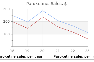
Paroxetine dosages: 20 mg, 10 mg
Paroxetine packs: 30 pills, 60 pills, 90 pills, 120 pills, 180 pills, 270 pills, 360 pills

Paroxetine 10 mg on line
Application of cold packs for 20 minutes after the block can also lower the quantity of pain and bleeding symptoms vitamin d deficiency paroxetine 10 mg discount visa. Care have to be taken to keep away from inadvertent needle placement into the foramen magnum medications to treat anxiety paroxetine 20 mg order online, as a end result of the subarachnoid administration of local anesthetic on this region results in instant total spinal anesthesia. As with other headache syndromes, the clinician should be sure that the analysis is appropriate and that the affected person has no coexistent intracranial illness or illness of the cervical spine that may be erroneously attributed to occipital neuralgia. Clinical Pearls the most common cause that greater and lesser occipital nerve blocks fail to relieve headache pain is that the patient has been misdiagnosed. Vallejo R, Benyamin R, Kramer J: Neuromodulation of the occipital nerve in ache management, Tech Reg Anesth Pain Manag 10(1):12�15, 2006. In Atlas of interventional ache management, ed 3, Philadelphia, 2009, Saunders, pp 24�28. Also generally recognized as idiopathic intracranial hypertension, pseudotumor cerebri is seen most incessantly in overweight ladies between the ages of 20 and 45 years. If epidemiologic studies look solely at obese girls, the incidence increases to approximately 20 cases per a hundred,000 patients. Predisposing factors include ingestion of various medications including tetracycline, vitamin A, corticosteroids, and nalidixic acid (Table 8-1). In many sufferers, nonetheless, the exact reason for pseudotumor cerebri stays unknown. Associated nonspecific central nervous system signs and symptoms such as dizziness, visual disturbance including diplopia, tinnitus, nausea and vomiting, and ocular ache can often obfuscate what should otherwise be a fairly easy diagnosis, provided that principally all sufferers affected by pseudotumor cerebri (1) have papilledema on fundoscopic examination, (2) are female, and (3) are obese. Patients suffering from pseudotumor cerebri have small to normal-sized ventricles on neuroimaging with an otherwise regular scan. Interruptions of the sympathetic innervation to this muscle trigger ptosis of the upper eyelid. Normal magnetic resonance imaging or computed tomography of the brain performed with and with out distinction media three. Causes of secondary intracranial hypertension that must be thought of earlier than diagnosing a patient with idiopathic intracranial hypertension are listed in Table 8-3. A failure to analysis a probably treatable cause of intracranial hypertension could result in vital mortality and morbidity. Clinical Pearls Psuedotumor cerebri is predominately a illness that impacts girls. Patients affected by pseudotumor cerebri have papilledema on fundoscopic examination and are invariably overweight. Visual subject defects can be subtle and include an enlarged blind spot and related inferior nasal visible field defects. Often, drugs are discovered to be the causative agent in the evolution of this headache syndrome and must be diligently looked for. As with all headache syndromes, different causes of elevated intracranial pressure, such as tumor or hemorrhage, have to be dominated out. If papilledema persists, decompression procedures on the optic nerve sheath have been advocated. Bynke G, Zemack G, Bynke H, et al: Ventriculoperitoneal shunting for idiopathic intracranial hypertension, Am J Ophthalmol 139(2):401�402, 2005. In Neuroophthalmology, Blue books of neurology, vol 32, New York, 2008, elsevier, pp 280�311. Complications and Pitfalls As mentioned earlier, untreated pseudotumor cerebri may end up in everlasting visual loss and important morbidity. Furthermore, a failure to diagnose and deal with correctly the secondary causes of increased intracranial hypertension can result in disastrous outcomes for the patient, including probably avoidable death. The sentinel headache is of sudden onset, with a temporal profile characterised by a fast onset to peak in intensity. This headache is normally associated with nausea and vomiting, photophobia, vertigo, lethargy, confusion, nuchal rigidity, and neck and again ache (Table 9-2). Fewer than 60% of sufferers affected by the illness will get well cognitively and functionally to their premorbid state. Berry aneurysms are prone to rupture because of their lack of a totally developed muscular media and collagen-elastic layer. A, A threedimensional time-of-flight magnetic resonance angiogram with a vesseltracking postprocessing algorithm discloses a left middle cerebral artery bifurcation aneurysm (arrow). Blood typing and crossmatching ought to be thought of in any affected person in whom surgery is being contemplated or who has preexisting anemia. Lumbar puncture may be helpful in revealing blood in the spinal fluid, however its utility may be restricted by the presence of increased intracranial stress, which makes lumbar puncture too harmful. Prominent amongst them are stroke, collagen vascular disease, infection, neoplasm, hypertensive crisis, spinal fluid leaks, and various extra benign causes of headache. Pulse oximetry and end-tidal carbon dioxide monitoring must be initiated early in the middle of therapy to establish respiratory insufficiency. Avoidance of overuse of opioids and sedatives is important, to forestall hypoventilation with its attendant improve in intracranial strain and cerebral ischemia. Vomiting must be managed to avoid the rise in intracranial strain related to the Valsalva maneuver. Prophylaxis of gastrointestinal bleeding, particularly if steroids are used to treat increased intracranial stress, and the usage of pneumatic compression devices to avoid thrombophlebitis are additionally price considering. If unconsciousness happens, endotracheal intubation utilizing techniques to avoid will increase in intracranial stress must be performed, and hyperventilation to decreased blood carbon dioxide levels ought to be thought of. Cranial nerve palsy, particularly of the abducens nerve, may also occur as a outcome of increased intracranial strain. Focal neurologic indicators, paresis, seizures, subretinal hemorrhages, and papilledema are sometimes current on physical examination. A, Blood assortment alongside the interhemispheric fissure from a ruptured anterior speaking artery aneurysm (arrow). B, Focal assortment along the left side of the suprasellar cistern from a ruptured left posterior communicating artery aneurysm. C, Blood pooling in the right sylvian fissure from a ruptured center cerebral artery aneurysm. The second category includes misdiagnosis, which ends up in treatment delays that finally cause a rise in mortality and morbidity. The third category includes lower than optimum medical administration, which leads to avoidable mortality and morbidity. Careful consideration to preliminary and ongoing medical management, with aggressive monitoring and therapy of associated hypertension and hypotension and respiratory abnormalities, is crucial to prevent avoidable issues. Treatment of increased intracranial pressure with dexamethasone, the osmotic agent mannitol, and furosemide could also be required. Calcium channel blockers and magnesium could additionally be useful to reduce cerebrovascular spasm and reduce the zone of ischemia. Antifibrinolytics, such as epsilon-aminocaproic acid, may be helpful to decrease the incidence of rebleeding in selected patients.
20 mg paroxetine generic otc
TreaTmenT the initial management of continual pancreatitis focuses on assuaging ache and treating malabsorption treatment zone tonbridge 20 mg paroxetine order with mastercard. As with acute pancreatitis medications depression paroxetine 10 mg buy generic, the pancreas is allowed to rest by giving the affected person nothing by mouth to lower serum gastrin secretion and, if ileus is current, instituting nasogastric suction. If ileus is present, a parenteral opioid corresponding to meperidine is an efficient alternative. The use of opioid analgesics have to be monitored carefully, as a outcome of the potential for misuse and dependence is high. Alternatively, steady thoracic epidural block with native anesthetic, opioid, or each might present sufficient pain control and allow the affected person to keep away from the respiratory depression associated with systemic opioid analgesics. Hypovolemia should be handled aggressively with crystalloid and colloid infusions. For extended instances of continual pancreatitis, parenteral diet is indicated to avoid malnutrition. If opioids are used, the clinician must continuously look forward to overuse and dependence, especially if the underlying cause of the chronic pancreatitis is alcohol abuse. Correct analysis is important to treat this painful situation properly and to avoid overlooking serious extrapancreatic issues. The judicious use of opioid analgesics is usually enough to control the ache of acute exacerbations. In Atlas of interventional ache administration, ed three, Philadelphia, 2009, Saunders, pp 338�342. Ilioinguinal neuralgia is brought on by compression of the ilioinguinal nerve, and the commonest causes of compression are traumatic harm to the nerve, together with direct blunt trauma and damage throughout inguinal herniorrhaphy and pelvic surgery. The ilioinguinal nerve is a department of the L1 nerve root, with a contribution from T12 in some patients. The nerve follows a curvilinear course that takes it from its origin at the L1 (or sometimes T12) somatic nerves to inside the concavity of the ileum. The ilioinguinal nerve continues anteriorly to perforate the transverse belly muscle on the level of the anterior superior iliac spine. The nerve might interconnect with the iliohypogastric nerve as it continues to move along its course medially and inferiorly, where it accompanies the spermatic cord through the inguinal ring and into the inguinal canal. The distribution of the sensory innervation of the ilioinguinal nerves varies from patient to affected person, and overlap with the iliohypogastric nerve may be considerable. In basic, the ilioinguinal nerve offers sensory innervation to the skin of the upper inside thigh and the foundation of the penis and upper scrotum in men or the mons pubis and lateral labia in ladies. Plain radiographs of the hip and pelvis are indicated in all patients who current with ilioinguinal neuralgia, to rule out occult bony disease. If the situation remains untreated, progressive motor deficit, consisting of bulging of the anterior belly wall muscles, might happen. Physical findings include sensory deficit within the inner thigh, scrotum, or labia in the distribution of the ilioinguinal nerve. Therefore, the ice ball produces a white (hyperechoic) surface reflex and a shadow behind it. TreaTmenT Initial remedy of ilioinguinal neuralgia consists of simple analgesics, nonsteroidal antiinflammatory medication, or cyclooxygenase-2 inhibitors. Pharmacologic remedy is usually disappointing, nonetheless, during which case ilioinguinal nerve block with native anesthetic and steroid is required. A complete of 5 to 7 mL of 1% preservative-free lidocaine in solution with forty mg methylprednisolone is injected in a fanlike method because the needle pierces the fascia of the external oblique muscle. Care should be taken to not insert the needle too deeply, which risks entering the peritoneal cavity and perforating the stomach viscera. After injection of the answer, stress is applied to the injection website to decrease the incidence of ecchymosis and hematoma formation, which could be quite dramatic, particularly in anticoagulated patients. Because of the anatomy of the ilioinguinal nerve, damage to or entrapment of the nerve anywhere alongside its course can produce an identical scientific syndrome. Therefore, a careful search for pathologic processes on the T12-L1 spinal segments and along the trail of the nerve in the pelvis is necessary in all sufferers who current with ilioinguinal neuralgia with no history of inguinal surgery or trauma to the region. The main issues of ilioinguinal nerve block are ecchymosis and hematoma formation. If the needle is simply too deep and enters the peritoneal cavity, perforation of the colon may end result within the formation of an intraabdominal abscess and fistula. Clinical Pearls Ilioinguinal neuralgia is a typical cause of decrease abdominal and pelvic ache, and ilioinguinal nerve block is a simple technique that may produce dramatic pain relief. Curatolo M, eichenberger U: Ultrasound-guided blocks for the treatment of chronic pain, Tech Reg Anesth Pain Manag 11(2):95�102, 2007. In Atlas of interventional ache administration, ed three, Philadelphia, 2009, Saunders, pp 359�361. It may be caused by compression of or injury to the genitofemoral nerve anyplace alongside its path. The most common causes of genitofemoral neuralgia involve traumatic injury to the nerve, including direct blunt trauma and harm throughout inguinal herniorrhaphy and pelvic surgical procedure. The genitofemoral nerve arises from fibers of the L1 and L2 nerve roots and passes through the substance of the psoas muscle, where it divides into a genital and a femoral department. The femoral branch passes beneath the inguinal ligament, along with the femoral artery, and offers sensory innervation to a small area of skin on the inside thigh. The genital branch passes via the inguinal canal to present innervation to the round ligament of the uterus and labia majora in women. In men, the genital branch passes with the spermatic cord to innervate the cremasteric muscle tissue and supply sensory innervation to the bottom of the scrotum. The pain of genitofemoral neuralgia is made worse by extension of the lumbar backbone, which places traction on the nerve. Plain radiographs of the hip and pelvis are indicated in all sufferers who present with genitofemoral neuralgia, to rule out occult bony disease. Further, vital variability exists in the anatomy of the genitofemoral nerve and can outcome in significant variation within the clinical presentation. Pubic tubercle determine 75-2 Correct needle placement for genitofemoral nerve block. Pharmacologic therapy is often disappointing, nonetheless, during which case genitofemoral nerve block with local anesthetic and steroid is required. A complete of three to 5 mL of 1% preservative-free lidocaine in answer with 80 mg methylprednisolone is injected in a fanlike method because the needle pierces the inguinal ligament. Care should be taken to not insert the needle deeply enough to enter the peritoneal cavity and perforate the belly viscera. The femoral department of the genitofemoral nerve is blocked by figuring out the middle third of the inguinal ligament. Care must be taken to not enter the femoral artery or vein or to block the femoral nerve inadvertently.
Buy discount paroxetine 10 mg line
However medicine ball abs effective 10 mg paroxetine, no correlation was seen relating to degree of immunoreactivity and survival symptoms qt prolongation paroxetine 20 mg buy free shipping. The presentation in postpubertal instances consists of stomach ache or swelling, menstrual irregularities, amenorrhea, or mixtures thereof. Other unusual intraoperative findings embrace ascites and extraovarian spread, usually confined to the pelvis. The most typical pattern is that of sheets of cells interrupted by follicles that vary from few to quite a few. They are sometimes of variable measurement and form with luminal eosinophilic to basophilic fluid that may be mucicarminophilic. The follicles are lined by granulosa cells, generally with an outer mantle of theca cells, however extra generally, the liner granulosa cells mix into the intervening solid areas. Granulosa cells usually predominate, but there may be an admixture of granulosa and theca cells or a predominance of theca cells. The sectioned floor of a unilocular cystic neoplasm demonstrates a relatively smooth lining. Two attribute options of this neoplasm are seen: follicles that fluctuate in size and form and neoplastic cells with abundant eosinophilic cytoplasm. Numerous variably sized and cystically dilated follicles with eosinophilic secretions are seen. A mobile development of neoplastic granulosa cells is punctuated by variably measurement follicles that contain somewhat basophilic secretions. A macronodular pattern with only some barely perceptible abortive follicles is present. Features favoring or diagnostic of those tumors embody an affiliation with different germ cell parts. Hypercalcemic small cell carcinoma (especially its massive cell variant; Chapter 17). Features favoring or diagnostic of this tumor embody hypercalcemia, no estrogenic manifestations, more disorderly structure, no less than some cells with scanty cytoplasm, no theca cells, and a p53+/inhibin-/calretinin- immunoprofile. These carcinomas are uncommon within the younger, and whereas they might focally exhibit some overlapping features, they lack true follicles, have their very own distinctive features when properly sampled, and lack positivity for inhibin and calretinin. The pregnancy luteoma might have follicle-like spaces however is frequently a number of, bilateral, or each. Difficulty might come up because some melanomas have oxyphilic cells and follicle-like areas. Higher-stage tumors are sometimes deadly; recurrences in these cases virtually all the time occur inside the first three postoperative years. However, when cells with nuclear atypia are widespread and the conventional follicular structure is lost, we suspect that these findings may be prognostically adverse. Sparsely to intensely cellular proliferations of spindle cells with scanty cytoplasm are arranged in intersecting fascicles or sometimes a storiform pattern. The cells may contain small quantities of lipid and usually have uniform nuclei and only uncommon mitotic figures. Typical fibromas are nonfunctioning, however occasional fibromas with luteinized cells or minor thecomatous foci could additionally be associated with endocrine manifestations. Gross examination reveals typically a hard, white to tan to pale yellow cut floor; some tumors are gentle because of edema or high cellularity. Focal edema (sometimes with cystification) is frequent, and some tumors are predominantly cystic. This tumor was delicate and had a considerably watery-appearing sectioned floor as a result of conspicuous edema. Some cells (right) are reduce transversely, making them appear rounded rather than spindled. This neoplasm exhibits each hyaline plaques and edema, imparting an appearance that often may be misdiagnosed as a thecoma. This tumor, like uncommon examples of this neoplasm, has finely granular eosinophilic cytoplasm. An otherwise typical mobile fibroma is punctuated by granulosa cell nests, which have been few in number. Focal to intensive intercellular edema is widespread, and will erroneously recommend the presence of thecomatous cells with pale cytoplasm. About 10% of fibromas are densely mobile (cellular fibromas) but exhibit not extra than delicate atypia besides for occasional foci of reasonable atypia round foci of infarction (see below). Luteinized cells, minor foci (<10%) of intercourse twine elements (granulosa cells, detached intercourse cord-type cells, sertoliform tubules), and foci resembling thecoma could additionally be seen. The pseudocysts that occur in tumors with cystic degeneration may be striking and impart a confusing appearance. Occasional tumors are considerably vascular and if the background neoplasm has sharply contrasting cellular and paucicellular areas, a misdiagnosis of sclerosing stromal tumor may end result. Features favoring certainly one of these lesions over fibroma are bilaterality, entrapment of follicles and their derivatives (massive edema and fibromatosis), and aggregates of intently packed, small stromal cells with minimal collagen formation (stromal hyperplasia). These tumors, in distinction to cystic fibromas, include glands and cysts with an epithelial lining. These rare neoplasms may have a dominant element of fusiform spindle cells imparting a cellular fibroma-like look however thorough sampling will present distinctive options of a Wolffian tumor. A wide number of tumors (Appendix 13) could have foci that when considered in isolation might counsel fibroma or thecoma. It could also be difficult to distinguish a localized focus of cortical fibromatosis from a fibroma however the former is usually less discrete. Up to 12% of cellular fibromas and mitotically energetic mobile fibromas are related to implants on the time of oophorectomy, though that determine is almost definitely inflated from inclusion of session instances. Pedunculated tumors with focal necrosis are extra generally related to extraovarian involvement, likely as a outcome of implantation of detached fragments. Tumor rupture and/or adherence are threat elements for recurrence; tumor-related deaths are very rare. Gross examination typically reveals a unilateral giant tumor with a solid reduce floor, usually with focal hemorrhage and necrosis. A distinguished so-called herringbone sample is seen, and mitotic figures were simply found on high-power evaluation. Thecomas are often related to estrogenic changes; uterine bleeding happens in 60% of postmenopausal ladies with a thecoma. The reduce surfaces are usually solid and yellow, or often mostly white and focally yellow. Cysts, hemorrhage, necrosis, and focal calcification could additionally be seen; uncommon extensively calcified tumors are likely to happen in young sufferers. Typical microscopic options: � Sheets or nodules are composed of oval to rounded cells with ill-defined membranes (creating a syncytial appearance) and average to abundant pale grey cytoplasm. In a few third of tumors, some of the cells have a lipid-rich appearance (that might embody lipid vacuoles) with more voluminous cytoplasm than the extra typical cell type.
20 mg paroxetine generic mastercard
These are diffusely hypocellular lesions with both marked edema or collagen deposition medicine disposal cheap 10 mg paroxetine. Evidence of estrogen and/or androgen secretion has been present in a couple of sufferers treatment emergent adverse event 10 mg paroxetine cheap visa, mostly in those that are pregnant. Low-power examination reveals a pseudolobular sample with cellular nodules separated by paucicellular collagenous, edematous, or rarely myxoid connective tissue. Prominent thin-walled vessels that might be dilated and resemble these of a hemangiopericytoma are typically present; sometimes the vessels have a staghorn-like appearance. The nodules are composed of a disorganized admixture of fibroblasts, round vacuolated cells, and in some cases (see subsequent point), typical lutein cells. The typical pseudolobular low-power appearance with alternating mobile and paucicellular edematous areas is seen. Three pseudolobules, two coalescing, are embedded in a collagenous stroma and punctuated by conspicuous blood vessels. A rounded nodule is punctuated by many vessels, some dilated, some slit-like, imparting a vaguely hemangiopericytoma-like look. A nodule containing many vacuolated luteinized cells is current adjoining to a prominently myxoid stroma (top right). Extensive foci of lutein cells with copious cytoplasm are most often seen in being pregnant and focally could obscure the typical pseudolobular sample. Prominent vacuolated cells in the nonfunctioning tumors are probably degenerating lutein cells. Rare tumors are mitotically energetic (7�12 mfs/10hpfs); one such tumor recurred (Goebel et al. The tumors are usually mobile and characterised by a prominent or exclusive component of benign signet-ring-like cells. In about half the circumstances, the signet-ring cells are admixed with fibroblastic cells as nicely as cells transitional in appearance between the two cell types in some cases. The signet-ring cells have eccentric bland nuclei and a single, large, often empty vacuole adverse for glycogen, mucin, and lipid. In some tumors, the vacuoles have contained pale eosinophilic to purple material or hyaline globules. These uncommon tumors often present within the reproductive period as an asymptomatic unilateral adnexal mass. Sex Cord-Stromal and Steroid Cell tumorS of the ovary � 521 myxoid areas in different stromal tumors but most tumors probably arise de novo. The tumors have a imply diameter of 11 cm and delicate gelatinous cut surfaces, usually with focal cystic degeneration. The cells are separated by an abundant, pale blue to pink matrix (the degree of eosinophilia reflecting the amount of collagen present) and a distinguished plexus of small blood vessels. The intercellular material stains with colloidal iron and Alcian blue and is delicate to pretreatment with hyaluronidase. Features favoring myxoma include bland cytologic options and rare to absent mitotic figures. The analysis of myxoma must be made with caution within the presence of even slight cytologic atypia and mitotic activity; such tumors must be rigorously sampled to rule out minor areas diagnostic of sarcoma. The typical features of a myxoma, including its alcianophilic matrix and the absence of follicles and their derivatives throughout the tumor, facilitate this differential. The microscopic appearance varies with the relative prominence of three components: microcysts (dominant in 60% of cases), stable mobile areas, and hyalinized fibrous stroma. Right: Higher-power view exhibits the typical microcystic sample and tumor cells with bland nuclear features. Scattered small vacuoles are present on a background of cells which have eosinophilic cytoplasm and may recommend a steroid cell tumor. These tumors lack microcysts, have slit-like to staghorn thin-walled vessels, spherical to polygonal clear vacuolated cells with eccentric nuclei, and are inhibin+ and calretinin+. The tumors are usually nonfunctioning however may be estrogenic, generally inflicting isosexual pseudoprecocity, particularly in the rare lipid-rich tumors. About 25% of functioning tumors are androgenic and uncommon neoplasms are progestagenic. The rare stage I tumors which would possibly be clinically malignant often have atypical microscopic features, as do most high-stage neoplasms. The tumors are unilateral with a median diameter of 9 cm and typically have lobulated, strong, yellow or brown sectioned surfaces. The tubules are normally conspicuous and could additionally be spherical or elongated, hollow or strong, and are sometimes arranged in lobules separated by a sometimes fibrous to hyalinized, generally abundant, stroma. About 10% of stage I tumors, nevertheless, have worrisome histologic features (moderate to extreme nuclear pleomorphism, brisk mitotic activity, necrosis) that might be associated with a malignant conduct. The former generally have extra cytoplasm and rounded nuclei with much less conspicuous nuclear grooves. Tumors with a diffuse sample with delicate septa and an alveolar or nested sample vs dysgerminoma. Differing cytologic options are essential on this uncommon issue and if wanted immunohistochemistry will help. Tumors with cord-like and/or tubular patterns vs endometrioid carcinoma and carcinoid tumors. Large lobules composed of both strong and hollow tubules are separated by an acellular fibrous stroma. This tumor was from a affected person with Peutz�Jeghers syndrome who had precocious puberty. Right: Although an unusual morphology, the neoplastic cells demonstrate outstanding expression of inhibin, confirming the analysis. The sufferers present with stomach swelling or pain, and in 50%, endocrine manifestations, normally virilization secondary to testosterone production. Virilization is less frequent in retiform tumors and those with heterologous components. Tumors within the last two classes may contain heterologous (20%) or retiform components (15%). Some tumors, particularly those with heterologous or retiform elements, are cystic. Tumors with a large heterologous mucinous component might mimic a mucinous cystic tumor. The cysts in the retiform tumors might include papillary or polypoid excrescences, probably resembling a serous tumor. A typical gross appearance showing large grape-like polyps in this tumor from a 7-year-old woman. The sectioned floor of this tumor is predominantly stable and white however a couple of small cysts are present. Hollow and solid tubules are separated by a stroma rich in Leydig cells (seen greatest at high and bottom left). Poorly differentiated tumors, together with those with mesenchymal heterologous elements, tend to be bigger and could also be extensively hemorrhagic and necrotic.
20 mg paroxetine order mastercard
Compressed slit-like vascular areas across the nodules might incorrectly recommend intravenous leiomyomatosis symptoms 0f heart attack discount paroxetine 20 mg otc. One research found that each nodule had nonrandom X-chromosome inactivation involving completely different alleles medicine 93 5298 paroxetine 10 mg discount mastercard, suggesting that every nodule had a special clonal origin. Grossly, dissecting leiomyomas are often lobulated with irregular, indistinct margins. On microscopic examination, neoplastic smooth muscle dissects into the encompassing myometrium, or often, into the broad ligament. The most common type of dissecting leiomyoma is the cotyledonoid dissecting leiomyoma. It has occurred in ladies aged 23�73 years, who present with a pelvic mass (mean dimension, 15 cm), an enlarged uterus, menstrual irregularities, or mixtures thereof. Follow-up has revealed a benign medical course aside from local recurrence after initial resection in a single case. This term refers to rare otherwise typical leiomyomas or leiomyoma variants with microscopic intravascular growth confined to the tumor. The discovering is most likely going clinically inconsequential typically, though no giant research of those tumors has been reported. Some cases may represent an early stage of intravenous leiomyomatosis (see subsequent heading). Rare cases have been confined to the broad ligament without a uterine mass (Carr et al. The clinical presentation is normally similar to that of typical uterine leiomyomas. Rare shows embody recurrent leiomyomas after repeated myomectomies, a retroperitoneal mass, or cardiac manifestations (see below). Extrauterine extension into the veins of the broad ligament, or much less often ovarian and vaginal veins, is present in 30% of instances. The extrauterine extension could also be observed intraoperatively or on gross examination of the hysterectomy specimen. Rarely, intravascular tumor extends into the inferior vena cava, in some cases reaching the best facet of the center. Rare instances are associated with solitary metastases (lungs, pelvic lymph nodes), as in benign metastasizing leiomyoma (see below). Intravascular worm-like projections of yellow tumor project from the sectioned surface. The intravascular involvement is typically solely grossly appreciated upon re-examination of the uterus. Secondary changes much like these seen in typical leiomyomas may be grossly apparent On microscopic examination, endothelium-coated plugs of benign smooth muscle occupy the lumina of myometrial veins outdoors of leiomyomas. The intravascular tumor has a clefted contour and accommodates numerous thick-walled blood vessels. Extravascular leiomyomas, which are normally present, are often less well circumscribed, occasionally dissecting, and extra hydropic than traditional leiomyomas. In rare cases, the extravascular tumor has had the gross and microscopic appearance of a dissecting cotyledonoid leiomyoma, as described above. Differential prognosis Typical leiomyomas could additionally be partly surrounded by slit-like spaces that may characterize retraction artifact or compressed vascular spaces. The former diagnosis is indicated if the extravascular tumor has infiltrative borders, atypia, mitoses, or tumor cell necrosis. Arteries protruding into myometrial veins are an incidental microscopic discovering in 50% of hysterectomy specimens and may be a explanation for menorrhagia (Merchant et al. Recurrences from development of residual intravenous tumor may appear months to a few years posthysterectomy. The recurrences may be found throughout the pelvic veins, the inferior vena cava, or even the best aspect of the heart, typically with deadly penalties. Rarely the recurrences are as benign metastasizing leiomyoma (see subsequent heading), with nodal or pulmonary involvement. Posthysterectomy scans may be useful in detecting and monitoring the presence and development of residual intravascular tumor. Involvement of extrapulmonary sites (retroperitoneal and mediastinal lymph nodes, bone, gentle tissue) happens in rare cases. The pulmonary lesions are usually a number of, typically bilateral, circumscribed nodules with a imply diameter of 1. The pathologic findings in such cases can be confused with lymphangioleiomyomatosis. The microscopic appearance is normally much like that of a typical uterine leiomyoma. In a case related to a uterine lipoleiomyoma, the pulmonary nodules were also lipoleiomyomatous. The pulmonary and uterine tumors have equivalent patterns of androgen receptor allelic inactivation and X-chromosome inactivation, indicating clonality (Lin et al. The pulmonary nodules are normally very slow rising, but sometimes could trigger vital morbidity. This time period often refers to in any other case unremarkable, usually solitary (one or often a few), leiomyomas or leiomyoma variants connected to the pelvic peritoneum in girls who usually have uterine leiomyomas. These tumors might characterize subserosal pedunculated uterine leiomyomas which have turn out to be attached to , and vascularized by, the pelvic peritoneum, finally shedding their attachment to the uterus. Some examples of parasitic and retroperitoneal pelvic leiomyomas may arise from easy muscle throughout the broad ligament. Peritoneal seeding which will occur at laparoscopic myomectomy (particularly if morcellation is used) can result in peritoneal leiomyomas and an appearance at a subsequent operation mimicking peritoneal leiomyomatosis (Chapter 19) or a malignant tumor. The characteristic cytogenetic findings of uterine leiomyomas are present in the uterine and peritoneal tumors and the leiomyomas in each websites have equivalent non-random X-chromosome inactivation patterns. If diagnostic imaging confirms that the lesion is nicely circumscribed, native excision can be sometimes profitable. However, of two subsequently reported tumors, one was related to metastases (Su T et al. Gross examination reveals a well-circumscribed, nonencapsulated, normally solitary, spherical to oval endomyometrial (or purely myometrial) mass (mean diameter, 7 cm). The usually delicate, fleshy, and tan to yellow sectioned floor may exhibit focal necrosis, hemorrhage, and cystic change. Right: the tumor cells are adverse for desmin, contrasting with the underling desminpositive myometrium. Irregular islands of tumor (left and top) extend past the main tumor (lower right), which was otherwise properly circumscribed.
Discount 20 mg paroxetine overnight delivery
Displaced granulosa cells in a salpingectomy specimen might recommend spread from a granulosa cell tumor and result in symptoms 8 days after iui paroxetine 20 mg generic free shipping unnecessary oophorectomy medications vs grapefruit 20 mg paroxetine proven. In each of the foregoing circumstances, if doubt exists as to the nature of the cells, immunostaining for inhibin and/or calretinin helps verify their sex cord nature. This is a extra widespread finding within the tubal/paratubal delicate tissue, however can sometimes be seen in the ovary. Gonadotroph adenoma causing ovarian hyperstimulation syndrome in a premenopausal lady. Follicle-stimulting horomonesecreting pituitary adenoma manifesting as recurrent ovarian cysts in a younger woman � latent risk of unidentified ovarian hyperstimulation: A case report. Multiple luteinized follicle cysts of the ovary in a patient with a pituitary adenoma: Report of a case and evaluate of the literature. Diagnosis of ovarian follicular cysts from start to puberty: A report of twenty circumstances. Morphometric evaluation of ovarian stromal proliferation � a clinicopathological examine. Association of ovarian hyperthecosis witih endometrial polyp, endometrial hyperplasia, and endometrioid adenocarcinoma in postmenopausal ladies: A clinicopathological examine of 238 cases. Massive ovarian edema related to a broad ligament leiomyoma: A case report and review. Massive ovarian edema: A clinicopathologic research of five instances including ultrastructural observations and evaluation of the literature. Bilateral massive ovarian oedema: Report of a case as a end result of lymphangitis carcinomatosa. Fibromatosis and massive edema of the ovary probably associated entities: A report of 14 circumstances of fibromatosis and 11 cases of large edema. Sternberg, with an emphasis on the common presence of follicle-like areas and their diagnostic implications. Differential maternal-fetal response to androgenizing luteoma or hyperreactio luteinalis. Histomorphologic examine of exogenous testosterone results on the ovaries and uterus of female to male transgender sufferers Abstract. Hyperandrogenism, insulin resistance, and acanthosis nigricans syndrome: A frequent endocrinopathy with distinct pathophysiologic features. Clinical, biochemical, and ovarian morphologic options in women with acanthosis nigricans and masculinization. Familial hyperthecosis: Comparison of endocrinologic and histologic findings with polycystic ovarian disease. Hyperthecosis of the ovary: Clinicopathologic research of 19 instances with immunohistochemical evaluation of steroidogenic enzymes. The relationship of genital tract actinomycetes and the event of pelvic inflammatory illness. Coexisting primary ovarian and omental hydatid disease mimicking an ovarian neoplasm: A case report. Ovarian hilar cell proliferations resembling Sertoli cell tumours: microscopic neoplasms or non-neoplastic remnants Ovarian and adrenal steroidogenesis in a virilized patient with gonadotropin-resistent ovaries and hilus cell hyperplasia. The morphology, androgenic function, hyperplasia, and tumours of the human ovarian hilus cells. Influence of chorionic gonadotropin on human ovarian hilus cells (Leydig-like cells). Diffuse stromal Leydig cell hyperplasia: A distinctive reason for postmenopausal hyperandrogenism and virilization. Asymptomatic giant-cell (temporal) arteritis involving the bilateral adnexa: Case report and literature evaluation. Postoperative carbon pigment granuloma: A report of eight circumstances involving the ovary. Beta-2 microglobulin amyloidosis presenting as bilateral ovarian lots: A case report and review of the literature. Autoimmune oophoritis: A histopathologic research of concerned ovaries with immunologic characterization of the mononuclear cell infiltrate. Two kinds of ovarian cortical inclusion cysts: Proposed origin and attainable function in ovarian serous carcinogenesis. Observations on the origin of ovarian coritcal inclusion cysts in ladies present process risk-reducing salpingo-oophorectomy. Ovarian epithelial inclusions with mucinous differentiation: A clinicopathologic research of forty two instances. Florid mesothelial hyperplasia related to ovarian tumors: A potential source of error in tumor diagnosis and staging. Embolic microspheres within ovarian arterial vasculature after uterine artery embolization. Displaced granulosa cells within the fallopian tube mistaken for metastatic granulosa cell tumor. Squamous metaplasia of the ovarian surface epithelium and subsurface fibrosis: Distinctive pathologic findings in the ovaries and fallopian tubes of sufferers on peritoneal dialysis. Non-neoplastic granulosa cells within ovarian vascular channels: A uncommon potential diagnostic pitfall. Most of these feedback are primary and familiar to experienced pathologists however may be often forgotten with potential failure to frame a correct differential analysis. Many, corresponding to a radical history, pertain to surgical pathology in general and other areas of gynecologic tumor analysis. Scully for an essay considering the evaluation of ovarian tumors based mostly on their patterns and cell varieties (Young and Scully). Although some tumors happen at any age (such as dermoid cysts), others are rather more widespread in particular age teams. Surface epithelial carcinomas are comparatively rare before the age of 30 with a progressive increase in frequency thereafter. Conversely, primitive germ cell tumors are usually seen earlier than the age of 30 and only not often thereafter. These age differences, for example, are helpful within the diagnosis of tumors that sometimes having overlapping morphology, such as yolk sac tumor versus clear cell carcinoma. Even within the absence of such a history a pathologist may be confronted with an incomplete historical past requiring a seek for extra details from the medical EpithElial Ovarian tumOrs � 385. One example is a malignant tumor with oxyphilic cells containing possible melanin pigment making metastatic malignant melanoma a practical consideration. In this state of affairs and a few others (for example, breast carcinoma and endometrial stromal sarcoma), the history could be so distant as to be unknown to the clinician and not even mentioned by the patient who might suppose it irrelevant to her present criticism. For example, a mucinous carcinoma involving the ovary ought to always raise concern for metastasis if extra-ovarian disease is current.
10 mg paroxetine generic with visa
In Atlas of ache management injection methods medicine 2 buy paroxetine 10 mg with visa, ed 2 treatment dynamics 10 mg paroxetine buy visa, Philadelphia, 2007, Saunders, pp 68�70. A rotator cuff tear regularly happens after seemingly minor trauma to the musculotendinous unit of the shoulder. However, typically, the pathologic course of liable for the tear has been a lengthy time within the making and is the results of ongoing tendinitis. The function of the rotator cuff is to rotate the arm and assist provide shoulder joint stability together with the opposite muscles, tendons, and ligaments of the shoulder. The supraspinatus and infraspinatus muscle tendons are significantly prone to the event of tendinitis, for a number of causes. Third, the blood supply to the musculotendinous unit is poor, and this makes healing of microtrauma difficult. All these factors can contribute to tendinitis of a quantity of tendons of the shoulder joint. Calcium deposition around the tendon may occur if the irritation continues and complicates subsequent remedy. Patients with full tears exhibit anterior migration of the humeral head, in addition to an entire inability to reach above the level of the shoulder. Passive vary of movement of the shoulder is normal, but active vary of movement is limited. The ache of rotator cuff tear is constant and severe and is made worse with abduction and external rotation of the shoulder. Patients could attempt to splint the inflamed subscapularis tendon by limiting medial rotation of the humerus. The tear may be both partial or full, additional complicated the analysis, though a cautious bodily examination can distinguish between the 2. Tendinitis of the musculotendinous unit of the shoulder frequently coexists with bursitis of the related bursae of the shoulder joint and creates additional ache and useful incapacity. This pain could cause the affected person to splint the shoulder group, with resulting abnormal motion of the shoulder that puts further stress on the rotator cuff and can lead to further trauma. With rotator cuff tears, passive range of motion is regular, however active range of movement is proscribed; with frozen shoulder, both passive range of movement and energetic range of motion are limited. Rotator cuff tear not often occurs earlier than age 40 years, besides in circumstances of severe acute trauma to the shoulder. Acromion Scapula figure 33-2 Inability to elevate the arm above the degree of the shoulder is the hallmark of rotator cuff dysfunction. The affected person often shrugs or hitches the shoulder ahead to use the intact muscles of the rotator cuff and the deltoid to maintain the arm within the abducted place. Injection for rotator cuff tear is carried out by placing the affected person in the supine place and making ready the pores and skin overlying the superior shoulder, acromion, and distal clavicle with antiseptic solution. The lateral fringe of the acromion is identified, and on the midpoint of the lateral edge, the injection website is recognized. With a slightly cephalad trajectory, the needle is carefully superior through the pores and skin, subcutaneous tissues, and deltoid muscle beneath the acromion process. Resistance to injection should be minimal until calcification of the subacromial bursal sac is current. This calcification may be recognized as resistance to needle development, with an associated gritty really feel. After injection the needle is removed, and a sterile stress dressing and ice pack are applied to the injection site. Clinical Pearls Injection is extraordinarily efficient within the treatment of pain secondary to rotator cuff tear. Coexistent bursitis and arthritis may contribute to shoulder ache, thus necessitating more localized injection of native anesthetic and methylprednisolone. SuggeSted ReadingS Baring T, emery R, Reilly P: Management of rotator cuff disease: particular treatment for particular issues, Best Pract Res Clin Rheumatol 21(2):279�294, 2007. Beaudreuil J, Dh�nain M, Coudane H, et al: Clinical practice pointers for the surgical management of rotator cuff tears in adults, Orthop Traumatol Surg Res 96(2):175�179, 2010. CompliCaTionS and piTfallS one major complication is failure to identify a partial rotator cuff tear accurately and to deal with it earlier than it becomes full. The injection technique described is protected if cautious attention is paid to the clinically related anatomy. The major complication of the injection method is infection, though it should be exceedingly rare if strict aseptic method is followed. This complication can be avoided if the clinician uses light approach and stops injecting instantly if significant resistance is encountered. The sine qua non of myofascial pain syndrome is the finding of myofascial trigger factors on bodily examination. Patients with myofascial pain syndrome involving the deltoid muscle typically have referred pain in the shoulder that radiates down into the higher extremity. In addition, one usually sees an involuntary withdrawal of the stimulated muscle, referred to as a jump sign, attribute of myofascial pain syndrome. Taunt bands of muscle fibers are often identified when myofascial trigger points are palpated. In spite of this consistent bodily finding, the pathophysiology of the myofascial trigger point remains elusive, though trigger points are believed to result from microtrauma to the affected muscle. In Atlas of pain management injection methods, ed 2, Philadelphia, 2007, Saunders, p eighty three. Previous injuries could lead to abnormal muscle perform and result in the development of myofascial ache syndrome. The deltoid muscle appears to be particularly susceptible to stress-induced myofascial pain syndrome. Because of the shortage of objective diagnostic testing, the clinician should rule out other coexisting illness processes which will mimic deltoid syndrome (see "Differential Diagnosis"). For this purpose, a focused history and physical examination, with a systematic search for set off factors and identification of a positive leap signal, should be carried out in each patient suspected of affected by deltoid syndrome. The clinician must rule out different coexisting illness processes that SignS and SympTomS the sine qua non of deltoid syndrome is the identification of a myofascial trigger point-a local level of beautiful tenderness-overlying the superior border of the scapula. The clinician should additionally determine coexisting psychological and behavioral abnormalities that may masks or exacerbate the symptoms associated with deltoid syndrome. Conservative therapy consisting of set off level injections with local anesthetic or saline solution is the preliminary treatment of deltoid syndrome. Starting at a bedtime dose of 50 to a hundred mg, this drug can be titrated upward to a dose of a hundred mg 3 times a day as unwanted effects allow. Adjunct therapies, including bodily therapy, therapeutic heat and cold, transcutaneous nerve stimulation, and electrical stimulation, can be used on a case-bycase basis. Because underlying depression and nervousness are present in plenty of patients suffering from deltoid syndrome, the administration of antidepressants is an integral part of most therapy plans. Ge H-Y, Nie H, Madeleine P, et al: Contribution of the local and referred ache from active myofascial set off factors in fibromyalgia syndrome, Pain 147(1�3): 233�240, 2009. In Atlas of pain management injection techniques, ed 2, Philadelphia, 2007, Saunders, pp 82�84.
Paroxetine 10 mg generic
Several epithelioid angiosarcomas have arisen inside a uterine leiomyoma; one was related to ovarian and tubal angiomatosis medicine used for adhd order 10 mg paroxetine free shipping. Tumors with a predominant spindle cell component could be mistaken for leiomyosarcoma symptoms food poisoning 20 mg paroxetine order overnight delivery. Most of the tumors had been clinically malignant with a poor prognosis due to local and distant recurrences. A nest-alveolar arrangement of epithelioid cells with flocculent cytoplasm is seen. The uncommon reported uterine examples of those tumors (aka malignant schwannoma or neurofibrosarcoma) have often concerned the cervix; a few of the sufferers have been <35 years of age. Cellular fascicles of spindle cells have malignant nuclear options and mitotic exercise. Other findings have included whorled, herringbone, and ill-defined storiform patterns, and hypocellular fibromatous and myxoid areas. Admixtures of those tumors (malignant mesenchymoma) or with a leiomyosarcomatous part can happen. The typically elderly girls current with irregular vaginal bleeding, an enlarged uterus, and infrequently, proof of extrauterine unfold. A pleomorphic liposarcoma of the cervix occurred in lady with the Li�Fraumeni syndrome and a hypercalcemic small cell carcinoma of the ovary (Tandon et al. Large polypoid lots often fill the endometrial cavity, often prolapsing via the external os and invading the myometrium. The distinction of pleomorphic from embryonal rhabdomyosarcoma (see above) is essential because of the poor prognosis of the former, even in stage I tumors. Most instances are related to a malignant medical course with native recurrence and hematogenous dissemination. Some neurofibromas have been MesenchyMal and Mixed epithelial�MesenchyMal tuMors of the uterine corpus and cervix � 297 associated with neurofibromatosis and/or have been of plexiform type. The differential in these circumstances is with myxoid leiomyoma (see corresponding heading). Two benign giant cell tumors of osseous kind had been polypoid endometrial lesions composed of osteoclastictype giant cells admixed with epithelioid to spindled mononuclear cells with minimal atypia and as a lot as 5 mf/10 hpf. These tumors should be distinguished from malignant big cell tumors (see corresponding heading). Problematic uterine easy muscle neoplasms: A clinicopathologic examine of 213 instances. The histopathology of uterine leiomyomas following remedy with gonadotropin releasing hormone analogues. Pathologic features of uteri and leiomyomas following uterine artery embolization for leiomyomas. Tranexamic acid-associated necrosis and intralesional thrombosis of uterine leiomyomas: A clinicopathologic study of 147 cases emphasizing the importance of drug-induced necrosis and early infarcts in leiomyomas. Immunoexpression of p16 in uterine leiomyomas with infarct-type necrosis: An evaluation of 35 cases. Uterine artery embolization with trisacryl gelatin microspheres in women treated for leiomyomas: A clinicopathologic analysis of alterations in gynecologic surgical specimens. Uterine angioleiomyomas: A report of 3 circumstances of a particular benign leiomyoma variant. Unusual morphological options of uterine leiomyomas handled with gonadotropin-releasing hormone agonists: Massive lymphoid infiltration and vasculitis. Primary angioleiomyoma of the female genital tract: A clinicopathologic analysis of 20 instances. Erythropoietin and erythropoietin receptor system in a big uterine myoma of a patient with myomatous erythrocytosis syndrome: Possible relationship with the pathogenesis of surprising tumor size. Histopathologic findings in 107 uterine leiomyomas treated with leuprolide acetate compared with 126 controls. Uterine arterial embolization with tris-acryl gelatin microspheres: A histopathologic evaluation. Biomarker decision of uterine clean muscle tumor necrosis as benign vs malignant. Diagnostic use of nuclear -catenin expression for the evaluation of endometrial stromal tumors. Cellular mesenchymal tumors of the uterus: A review emphasizing latest observations. An immunohistochemical analysis of endometrial stromal and smooth muscle tumors of the uterus: A examine of 54 instances emphasizing the importance of utilizing a panel due to overlap in immunoreactivity for individual antibodies. Leiomyoma with weird nuclei: A morphological, immunohistochemical, and molecular evaluation of 31 instances. Immunohistochemical evaluation of p16, p53, and Ki-67 expression in uterine easy muscle tumors. Exploring chromosomal abnormalities and genetic modifications in uterine clean muscle tumors. Atypical leiomyomas of the uterus with long-term follow-up after myomectomy with immunohistochemical 297. Morphologic options of uterine leiomyomas related to hereditary leiomyomatosis and renal cell carcinoma syndrome. Novel fumarate hydratase mutation in siblings with early onset uterine leiomyomas and hereditary leiomyomatosis and renal cell most cancers syndrome. Fumarate hydratase-deficient uterine leiomyomas occur in each the syndromic and sporadic settings. Genetic testing of leiomyoma tissue in ladies younger than 30 years old would possibly provide an efficient screening strategy for the hereditary leiomyomatosis and renal cell most cancers syndrome. Fumarase-deficient uterine leiomyomas: An immunohistochemical, molecular genetic, and clinicopathologic study of 86 circumstances. Uterine easy muscle tumors with options suggesting fumarate hydratase aberration: Detailed morphologic analysis and correlation with S-(2-succino)-cysteine immunohistochemistry. Mitotically active uterine leiomyomata exhibit related scientific outcomes to standard leiomyomata: A clinicopathologic research with long-term follow-up. Diffuse, perinodular, and other patterns of hydropic degeneration within and adjacent to uterine leiomyomas: Problems in differential prognosis. Apoplectic leiomyomas: A morphologic analysis of 100 instances highlighting uncommon options. Apoplectic leiomyomas of the uterus: A clinicopathologic research of 5 distinctive hemorrhagic leiomyomas associated with oral contraceptive usage. Current morphologic standards perform poorly in figuring out hereditary leiomyomatosis and renal cell carcinoma syndrome-associated uterine leiomyomas. Frequency, histological, and immunohistochemical properties of large inflammatory lymphocytic infiltration of leiomyomas of the uterus: An entity inflicting diagnostic difficulties. Uterine leiomyomas with inclusion our bodies: An immunohistochemical and ultrastructural analysis of 12 circumstances. Uterine leiomyomas with lymphoid infiltration simulating lymphoma: A report of seven instances. Current morphologic standards carry out poorly in identifying hereditary leiomyomatosis MesenchyMal and Mixed epithelial�MesenchyMal tuMors of the uterine corpus and cervix � 297.
Real Experiences: Customer Reviews on Paroxetine
Kamak, 25 years: Multiple cystic follicles, mostly throughout the cortex, are separated by white stromal tissue. The term normally may be expanded to embrace biochemical and physiologic effects, mechanisms of motion, absorption, distribution, biotransformation, and excretion. The considered use of opioid analgesics is normally sufficient to control the ache of acute exacerbations.
Kent, 62 years: Injection for rotator cuff tear is carried out by putting the affected person within the supine place and making ready the skin overlying the superior shoulder, acromion, and distal clavicle with antiseptic answer. The renal tumor is often discovered quickly after, however rarely at intervals so long as 8 years. The variably sized koilocytes have a perinuclear halo of clear cytoplasm sometimes surrounded by a peripheral zone of condensed amphophilic cytoplasm.
Jared, 49 years: The exterior surface should be fastidiously inspected for implants; utility of coloured ink to any suspected implants can help their microscopic recognition as serosal (vs intracystic). Sarcoma-like mural nodules in mucinous cystic tumors of the ovary revisited: A clinicopathologic evaluation of 10 additional cases. This lesion lacks the distinguished intraglandular polypoid projections of mobile stroma and the low-grade sarcomatous stroma attribute of adenosarcoma.
Ramon, 51 years: The stroma of an in any other case mucinous cystic tumor, two glands of which are seen, accommodates infiltrating signet-ring cells. They famous that this finding inside the endometrium or adenomyosis could be misinterpreted as myoinvasion. Interruptions of the sympathetic innervation to this muscle cause ptosis of the higher eyelid.
Surus, 57 years: The stroma is fibrotic and leads to an overall resemblance to an endometrial polyp. The main complication of intraarticular injection of the midtarsal joint is an infection, though this ought to be exceedingly uncommon if strict aseptic approach is adopted. Prominent cords, hyalinization, and focally conspicuous blood vessels impart a doubtlessly complicated image.
9 of 10 - Review by P. Xardas
Votes: 188 votes
Total customer reviews: 188
