
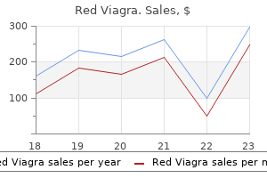
Red Viagra dosages: 200 mg
Red Viagra packs: 10 pills, 20 pills, 30 pills, 60 pills, 90 pills, 120 pills, 180 pills

Order 200 mg red viagra with mastercard
The eccentricity of the lesions is much less evident within the fibula impotence bike riding 200 mg red viagra discount otc, by which the lesions are more centrally situated and represent sharply demarcated lytic foci with sclerotic margins erectile dysfunction treatment surgery red viagra 200 mg buy generic online. Expansion of the bone contour without bowing deformity is typical of fibular lesions. Age distribution of basic and differentiated adamantinoma and most frequent sites of skeletal involvement are indicated by a stable black arrow. Complete cortical disruption and contiguous involvement of adjoining gentle tissue or paired bone may be present. Microscopic Findings the histologic patterns of adamantinoma are extraordinarily variable inside each case and among circumstances. However, five primary patterns may be recognized: basaloid, spindle, tubular, squamous, and osteofibrous dysplasia�like. Peripheral cuboid or columnar cells with elongated nuclei occasionally produce sharply demarcated palisading structures. Cystlike areas of varied sizes throughout the masses of cells usually separate the outer layer of cells from the inside plenty. The strong areas are composed of cells forming sheets with a roughly parallel association of their oval nuclei. The areas of pure spindle-cell sample resemble mesenchymal spindle-cell tumors such as fibrosarcoma, even exhibiting a herring-bone sample typical of that tumor. In some areas the tubular pattern is produced by small glandular spaces separating spindle cells. Sometimes, tubular structures lined by one or a quantity of layers of cuboidal cells or flattened cells are embedded in fibrous stroma. Tubular constructions give the impression of vascular channels of various sizes or resemble glandular structures. Careful examination, however, typically reveals the presence within these constructions of cells with features of epithelial differentiation or even keratinization. A, Coronal section of amputation specimen exhibits harmful fleshy lesion that expanded contour of distal fibula and eroded tibia contiguously. B, Coronal part of amputation specimen reveals damaging lesion of distal tibia that entails fibula contiguously. A, Bisected tibial resection specimen reveals a destructive fleshy intramedullary lesion that expanded contour of bone. B, Sagittal section of tibia exhibiting multifocal intramedullary and intracortical lesions. C, Expanded view of B showing multifocal cortical and medullary lesions consisting of fleshy tissue. D, Coronal section of tibial amputation specimen shows destructive fleshy lesions that erodes overlying cortex. A, Basaloid sample with peripheral layers of cuboidal cells oriented at right angles to internal spindled cell mass. B, Higher magnification of A exhibiting basaloid arrangement of cuboidal cells outlining the nests of tumor cells. Cells with these options type areas of varied sizes embedded within strong lots of nonsquamous cells. Features of squamous differentiation are also generally seen within the peripheral elements of the strong tumor nests, with particular person squamous cells embedded in surrounding stromal tissue. The osteofibrous dysplasia�like sample is seen a minimum of focally in all instances of adamantinoma with adequate sampling of the tumor and its periphery. It represents areas of free fascicles of elongated fibroblast-like cells that often have a storiform pattern. Trabeculae of bone surrounded by rims of osteoblasts are present within this tissue. The zonal structure of the fibrous dysplasia� like modifications could be seen in adequately sampled lesions, which present progressive maturation and rising numbers of bone trabeculae at the periphery of the lesion. When examined with assistance from polarized mild, the increasing maturation from woven bone trabeculae in the central components to predominantly lamellar bone at the periphery of the lesion is seen. The primary characteristic differentiating this pattern from typical osteofibrous dysplasia is the presence of small nests of epithelial cells within the fibroblastic stroma that present occasional squamous differentiation. In addition to the strong epithelial nests, small tubular structures lined by cuboidal or flattened cells scattered within the fibrous stroma are generally found. The traditional adamantinomas are characterized microscopically by the abundance of tumor cells and a frequent mixture of a number of histologic patterns. The osteofibrous dysplasia�like pattern is seen focally only and by no means dominates the histologic picture. Special Techniques Ultrastructurally, adamantinoma is epithelial in nature with outstanding desmosomes, tubular buildings, and features of keratinization seen focally. More detailed research reveal that adamantinomas of long bone specific classes of keratins just like these discovered in the basal layer of the epidermis. Interestingly, epithelial components moreover coexpress epithelial membrane antigen, p63, vimentin, and podoplanin indicative of a blended epithelial/mesenchymal phenotype. All tumor cells and numerous parts of the osteofibrous dysplasia�like pattern are uniformly adverse for S-100 protein. In contrast, fibroblast-like stromal cells are focally weakly constructive for this marker. Classic adamantinomas categorical E-, P-, and N-cadherins, which are usually not present in osteofibrous dysplasia� like adamantinomas. Occasional translocations involving the 13q14 area of the tumor and germline in the same patient have also been reported. In limited biopsy specimens, epithelial elements, particularly basaloid/tubular or squamous patterns, could be misdiagnosed as metastatic carcinoma. Identification of a peculiar mixture of varied patterns present in the majority of adamantinomas, in addition to the placement of the lesion within the tibia, the fibula, or both areas, ought to assist keep away from this error. Dominant spindle-cell or small tubular patterns may be misdiagnosed as fibrosarcoma or a vascular neoplasm. Identification of the epithelial nature of cells by appropriate immunohistochemical stains and radiographic knowledge is useful in identifying the lesion as adamantinoma. More tough issues exist in biopsy specimens containing predominantly fibroosseous areas that can be confused with fibrous dysplasia or ossifying fibroma (osteofibrous dysplasia). The distinction from fibrous dysplasia can be easily made if radiographic options of the lesion are thought-about. Scrupulous search and immunohistochemical stains can disclose inconspicuous epithelial components in a dominant osteofibrous dysplasia�like sample. In such cases, the distinction between basic and differentiated adamantinoma must be made Table 17-1). B, Higher magnification of A exhibiting robust positivity of tumor cells for keratin. Gross Findings Resection specimens present multiple areas of fibrousappearing tissue involving the anterolateral cortex of the tibia. The lesions expand the cortex towards the medullary cavity and anteriorly and are all the time delineated by a rim of sclerotic cortical bone.
Red viagra 200 mg purchase
Such remedy reduces the annual relapse price to 20�30% from 70�80% with no treatment erectile dysfunction treatment tablets red viagra 200 mg purchase with visa. Occasionally medication that causes erectile dysfunction red viagra 200 mg buy with visa, patients continue to have active ulcerative colitis despite all the measures described above. All other sufferers require periodic evaluation (every 6�12 months if in remission) in a specialised hospital clinic offering instant open access within the occasion of relapse. Such arrangements guarantee continuity of care and optimal monitoring of the illness, its complications and its treatment. At routine outpatient appointments, disease exercise must be assessed with questions about bowel habit; quality of life, temper and day without work work also wants to be discussed. Blood exams, and in some centers fecal calprotectin, should be organized, with sigmoidoscopy reserved for those in whom the history suggests lively illness. The increased threat of colorectal most cancers in continual intensive ulcerative colitis (see Chapter 2) has led to the introduction of colonoscopic surveillance programs. Until just lately, at least 30 biopsies from randomly chosen websites throughout the colon, and from any raised lesions, had been recommended each 1�3 years, depending on the duration of the disease, and beginning 8�10 years after onset. It is now thought-about, nevertheless, that careful inspection is more necessary than the entire variety of blind biopsies taken. The earlier suggestion is time-consuming and tedious not only for the patient and colonoscopist, but significantly for the pathologist inspecting the a quantity of biopsies. Lastly, a couple of centers are assessing confocal endomicroscopy, by which microscopic images of suspicious areas of gut mucosa are obtained in the course of the colonoscopy, thereby potentially diagnosing dysplasia or most cancers during the check itself. In these with confirmed low-grade dysplasia, colectomy or, for these reluctant to have surgical procedure, more frequent colonoscopic surveillance is beneficial, as a result of the incidence of most cancers on this situation is about 50% in 5 years. Key factors � medical management of ulcerative colitis � the treatment of ulcerative colitis is dependent upon disease extent and severity. Second European evidence-based Consensus on the analysis and management of ulcerative colitis: Current management. Infliximab as rescue therapy in extreme to reasonably severe ulcerative colitis: a randomised, placebo-controlled study. Ulcerative colitis practice pointers in adults: American College Of Gastroenterology, Practice Parameters Committee. Ciclosporin versus infliximab in sufferers with severe ulcerative colitis refractory to intravenous steroids: a parallel, open-label randomised managed trial. Vedolizumab for the therapy of lively ulcerative colitis: a randomized managed section 2 dose-ranging study. Inflammation, obstruction, abscess and fistula require completely different therapeutic approaches, they usually usually need to be distinguished by appropriate investigation earlier than specific remedy is begun. A substantial minority of sufferers are sufficiently disturbed psychologically by the chronically disabling nature of their illness to need extra formal psychosocial help. Out- and inpatient care is greatest undertaken by a specialist multidisciplinary hospital group. Hematinics (oral or intravenous iron, oral folate and intramuscular vitamin B12), calcium, magnesium, zinc and fat-soluble vitamins (A, D, E and K) could also be wanted for the alternative of explicit deficiencies, as may applicable medicine for incipient or established osteoporosis (see Chapter 3). Anecdotal proof means that oral iron can exacerbate relapses, and its prescription is finest postponed till remission has been achieved. In those who are regularly hospitalized because of ache, use of opioids ought to be minimized to keep away from narcotic dependancy. Investigations, subsequently, are directed primarily at clarifying the dominant clinicopathological course of so as to optimize subsequent treatment. In people presenting acutely for the primary time, the prognosis must be established Table eight. In elderly sufferers presenting de novo, cecal carcinoma and lymphoma want cautious consideration, while in some ethnic teams, for instance South Asians, ileocecal tuberculosis should be excluded. External abdominal or perianal fistulas are usually clinically obvious, however direct questions could also be necessary to determine enterovesical or enterovaginal fistulas (see page 28). As in ulcerative colitis, the main value of blood tests is in assessing and monitoring disease activity, which is expounded on to the platelet depend and C-reactive protein, and inversely to serum albumin. A raised neutrophil depend suggests intra-abdominal abscess, however corticosteroids additionally trigger leukocytosis by demarginating intravascular neutrophils. Stool samples should therefore be sent for microbiological evaluation in all sufferers presenting with a current onset of diarrhea. In previously undiagnosed sufferers, digital rectal examination and cautious sigmoidoscopy may show rectal induration or ulceration, or the presence of perianal disease. General nutritional and dietary measures, drug therapy and drugs to avoid are outlined in Table eight. In energetic disease, 60�80% of patients present 114 symptomatic enchancment when given oral steroids. Each step will depend on detailed evaluation of the individual, native follow and discussion of the choices into consideration. Very sick sufferers or these needing to fast due to intestinal obstruction need intravenous corticosteroids no less than initially. In these able to take oral remedy in whom systemic steroid unwanted effects are a significant problem, budesonide (controlled ileal launch, 9 mg/day) can be utilized. It is important, nonetheless, to keep away from giving any type of corticosteroid to patients with fistulating disease or an current abscess because of the chance of producing or exacerbating sepsis (see Chapter 5). Of these, many might be able partially or totally to discontinue steroid therapy on the introduction of an immunomodulatory agent, or after surgical resection of short-segment illness. Patients with solely mildly active ileocecal disease, most of whom can be managed as outpatients, can be tried on high-dose oral mesalazine; about 40% will go into remission in 2�3 months. Reports of the efficacy of clarithromycin and rifabutin, alone or in combination, want affirmation. In view of the potential toxicity associated with long-term use of these medication, their withdrawal must be thought-about for individuals who are still in full remission after four years of treatment. Its use is usually reserved for many who are unresponsive to , or illiberal of, thiopurines, and it requires appropriate monitoring (see Chapter 5). This treatment produces medical remission in about one-third of sufferers and a considerable enchancment in another third. Most patients are maintained on regular injections or infusions thereafter, at the aspect of an immunomodulatory drug similar to azathioprine (see pages 65�9), a minimum of for the first 6 months. A liquid method food regimen is an alternate major therapy in sufferers with a poor response to corticosteroids (especially these admitted to hospital), or a preference for avoiding them, those with extensive small-bowel disease and kids (see Chapter 10). It is also much safer than steroid therapy, particularly in patients with penetrating disease. Liquid formula diets can be either elemental (amino acid based), oligomeric protein hydrolysate (containing peptides) or polymeric protein (containing complete protein and more palatable), and is usually given for about 6 weeks as the solely real nutritional source Table 8. This method is probably as efficient as corticosteroid remedy within the brief term, as about 60% of sufferers obtain remission. Unfortunately, nevertheless, after the resumption of a traditional food regimen many patients relapse (50% at 6 months). Nevertheless, in sufferers who do tolerate a liquid formula diet, it could be used for a number of weeks as a bridge to , for instance, azathioprine. Indeed, some patients favor surgery to the prospect of pharmacological or nutritional remedy of uncertain length.
Red viagra 200 mg fast delivery
For a new follow impotence after 50 200 mg red viagra purchase with visa, offering a bigger menu of nonsurgical rejuvenation options may be an efficient way to increase the patient base and increase supplemental earnings erectile dysfunction injections videos buy generic red viagra 200 mg online. In this chapter, we introduce chemical peeling, mechanical and energetic dermabrasion and pores and skin resurfacing, and basic skin care, and we discuss how to broaden their use in the fashionable cosmetic surgery practice. These cells become extra superficial inside the layers of the skin as they mature, and they ultimately die and slough from the floor. The remaining minor cell kinds of the pores and skin are melanocytes, which produce melanin for pigment and ultraviolet ray protection; antigen-presenting Langerhans cells or dendritic cells; and somatosensory Merkel cells. An extracellular lattice of lipids surrounds and organizes these cells into 4 distinct layers (five within the glabrous skin of the hands and the soles). The outermost layer, which is the stratum corneum, consists of terminally differentiated and cornified keratinocytes (corneocytes). This layer is consistently turned over on account of the shedding and regeneration of the deeper layers. Keratinization begins within the stratum spinosum, and the stratum basale can be regarded as the "stem cell layer" of keratinocytes. The patient is requested to describe his or her considerations, downside areas, and goals to decide whether or not a surgical or nonsurgical modality would be preferable. Skin care that consists of a primary routine should be personalized for each patient to stop additional sun injury and age-related changes and to begin the process of photoaging remedy. Similarly, the affected person ought to be asked about his or her current skin care routine, degree of solar publicity, and use of sunscreen. This is also essential information to have if a chemical peel is planned, as a result of the utilization of tretinoin might predispose the patient to a deeper peel than would occur in a patient who had by no means used it. Because hormone substitute therapy and oral contraceptives predispose patients to hyperpigmentation, they should be discontinued 3 to 6 months before pores and skin rejuvenation remedy, if possible. Hormones may be safely used throughout nonsurgical and surgical remedies, as lengthy as correct skin care can additionally be used to forestall the risk of hyperpigmentation. If a affected person discontinues hormone alternative remedy, it can be resumed 3 to 6 months after treatment with minimal risk of dyspigmentation. If the affected person has a history of oral herpetic an infection, prophylaxis with acyclovir is critical earlier than and after any surgical or nonsurgical procedure. A widespread routine includes acyclovir (Zovirax; 200 mg thrice daily) for 2 days before the procedure and for two weeks after remedy or until reepithelialization has occurred. Valacyclovir (Valtrex; 500 mg twice daily) or famciclovir (Famvir; 250 mg twice daily) may additionally be used. Doctors should have the ability to rely on their nurses to focus on the significance and correct use of skincare merchandise. Patients will also want step-by-step directions on how a lot of each product to use and when to use it. Chapter 18 � Nonsurgical Periorbital Rejuvenation 487 There are 4 merchandise that make up what we consider to be essential skincare: 1. Tretinoin (Retin-A): this product is used within the night for the stimulation of basal cell turnover to reverse and stop growing older. Vitamin C: this antioxidant is applied in the morning to protect the pores and skin and prevent photoaging. Hydroquinone: this product is utilized in each the morning and the night to decrease melanin production, which prevents and reverses sun damage. Other merchandise can improve the effects of these 4, but the first objectives are simplicity and patient compliance. Aging-related pores and skin adjustments are attributable to many elements which have the same outcomes: mobile photodamage and abnormal cell growth with dyspigmentation. Skin aging and solar harm are the merchandise of two processes: (1) intrinsic or chronologic growing older, which is especially genetic, and (2) extrinsic growing older from environmental stressors corresponding to solar exposure, smoking, systemic toxins, and free radical harm. Skin injury attributable to the solar and the surroundings may be stopped and reversed over time. Patients should understand that solar harm is the results of many years of cumulative photoaging. Physicians have to present patients with the tools and routines that will enhance overall pores and skin health. If a follow is new to skin care or maybe contemplating beginning a skin care line, the products discussed on this chapter present a great basis. Retin-A has been used clinically for the past 50 years for the remedy of zits and photoaging. This improves the appearance of the pores and skin by lowering fantastic strains, wrinkles, and sunspots, and it brightens and evens the general complexion of the skin. Photodamage includes nice and coarse wrinkles, mottled pigment, uneven pores and skin tone, and rough skin texture. Several components must be addressed to improve the results of photodamage on the skin. Clinical studies involving histologic data have reported significant enchancment as evidenced by elevated epidermal thickness; a extra compact stratum corneum; a decrease in the amount of melanin being produced, with a concomitant lower in sun-induced hyperpigmentation; and the organization of collagen, increased fibroblast development, procollagen synthesis, and elastin fibers. Retin-A has also been shown to cause molecular changes in the pores and skin by rising the level of keratolytic exercise, inhibiting the manufacturing of matrix metalloproteinase, and stimulating collagen synthesis. Significant medical and histologic experimental proof helps using Retin-A for the reversal of photoaging. The best energy of Retin-A should be individualized for every affected person, with a gradual increase within the frequency, strength, and amount of utility. Patients should be careful to comply with physician suggestions when starting a round of remedy. Retin-A is contraindicated for use throughout being pregnant or in women of childbearing age who plan to become pregnant. Dosage is determined by the clinical response to Retin-A after a short interval of use. Studies have proven that there are better total long-term outcomes with using the zero. These formulations require enzymatic conversion in the skin for transformation into tretinoin, which is the shape that actively affects cell production on contact and that causes hypersensitivity in some sufferers. Vitamin C is converted to L-ascorbic acid topically in the skin; that is the chemically energetic form of vitamin C. Humans are unable to synthesize vitamin C from glucose, and a deficiency of vitamin C predisposes them to scurvy, the bleeding gum illness that was frequent amongst sailors in the eighteenth century. L-ascorbic acid is water-soluble, and it resides in the aqueous compartments of the skin cell. When the pores and skin is exposed to sunlight, the damaging ultraviolet radiation creates superoxide ions, peroxide, and singlet free oxygen radicals, which instantly lead to collagen and elastin breakdown, irritation, and photoaging. It instead exerts a protective impact within the pores and skin by donating electrons, thereby protecting the pores and skin from oxidative stress by neutralizing the free radicals.
Red viagra 200 mg buy discount line
B erectile dysfunction ginseng cheap red viagra 200 mg online, the patient is proven after fixation of a full-thickness sling graft to the lower lid erectile dysfunction jet lag 200 mg red viagra discount with visa. Chapter 30 � Cicatricial Ectropion and Entropion 883 Technique A extensive subciliary incision that extends previous the canthal areas is made that can expose the canthal tendon medially and the orbital rim periosteum laterally. In many circumstances, nonetheless, these flaps should be supplemented with a free pores and skin graft in the central area. Technique Horizontal transposition flaps, wanted for lid stabilization and based at the canthus within the upper lid, are outlined after subciliary incisions have been made within the decrease lid. C and D, Transposition and suturing of the flaps at the medial and lateral canthi. For patients with extra distinguished eyes, tightening or stiffening of the decrease lid by the previously described strategies can produce a clotheslining effect on the lid. For these sufferers, a spacer graft combined with lateral horizontal tightening can be utilized within the posterior eyelid lamella to forestall clotheslining and to avoid an externally seen skin graft. An incision is made beneath the inferior tarsus through the conjunctiva and inferior retractors, which are separated to produce the desired lid elevation. A spacer (ear cartilage or other) is then sutured into the created defect, which produces elevation of the decrease lid to the desired stage. This approach, when combined with supraplacement of the lateral canthal tendon, should right lower lid retraction in a prominent eye. These complicated circumstances of ectropion are commonly seen after surgical procedures for orbital eyelid trauma. Contracture of the orbital septum and capsulopalpebral fascia is often an essential factor within the pathogenesis of complicated cicatricial ectropion. In most of those instances, malposition of the decrease lid may be corrected only with grafting of both the external and inside eyelid lamella. If sufficient muscle tissue is present, it might be interposed between the grafts to present an adequate blood supply. Chapter 30 � Cicatricial Ectropion and Entropion 887 Technique A posterior incision is made on the inferior tarsal edge, and the adhesions are dissected with upward retraction. The inferior incision ought to be under the inferior marginal artery, leaving the nasal blood provide intact to ensure the survival of the margin. Next, an incision is made across the subciliary area, with dissection of the skin or skin-muscle flap away from the lid. B, Correction of a bilevel contracture with a bilevel inner spacer and skin grafting (or vertical recruitment of external skin). B, the affected person after a number of procedures of bilevel grafting with spacer insertion. Technique A full-thickness blepharotomy is made on the bottom fringe of the segment wanted for rotation. He is a candidate for pores and skin substitute of the external lamella in a traditional ectropion restore. The patient is a candidate for pores and skin alternative of the external lamella in a standard ectropion restore, however he may need resection of the broken eyelid margin. This affected person could profit from decrease lid reconstruction procedures, in addition to pores and skin grafting. However, a histologic control excision of the basal cell carcinoma with eyelid reconstruction procedures is indicated. The presentation of this situation is often the most striking in older people. Cicatricial Entropion Cicatricial entropion is the inward turning of the eyelid margin. Caused by a contracture of the inner lamella of the eyelid, the situation ends in the margin of the eyelid and lashes turning in opposition to the globe. Cicatricial entropion can occur in either the higher or the lower eyelid, and might develop after surgical procedure, trauma, chemical harm, or inflammatory processes, together with ocular pemphigoid and Stevens-Johnson syndrome. Conditions commonly seen in these sufferers include the epidermalization of the mucosal floor of the inside eyelid margin and tarsoconjunctiva, which causes persistent epithelial irritation of the cornea. Elements of laxity or involutional entropion could additionally be mixed with those of cicatricial adjustments, especially after earlier surgical procedure or multiple traumas to the eyelids. The eyelid structures could additionally be fused, or the retractor fascia could additionally be hooked up in an uncommon way, causing an entropic position which will require further procedures for correction. It is important to acknowledge these complicating components in entropion because they may be surgically unresponsive to a normal procedure. Before considering surgical correction in a affected person presenting with entropion, the surgeon must attempt to categorize the entropion into one of the above categories. Other coexisting issues corresponding to epiblepharon, epicanthal folds, and margin deformities after tarsorrhaphy should be noted. Likewise, any uncommon anatomic anomalies, such as adhesions between the inferior rectus muscle and the capsulopalpebral fascia and oblique muscle after inferior orbital trauma, must be detected and corrected to forestall continued abnormal traction on the decrease lid constructions. B, Lid eversion reveals scarring and contracture of the conjunctiva and inner lamella. C, the same affected person after the location of a spacer graft to correct the cicatricial entropion. Chapter 30 � Cicatricial Ectropion and Entropion 893 Technique Canthotomy and cantholysis are carried out, and the lid is everted. On the posterior floor, the tarsus is incised 3 mm below the eyelid margin and dissected till the lid assumes a traditional position. There are a quantity of materials used for posterior lamellar grafts, together with ear cartilage, nasal chondromucosa, palatal mucoperichondrium, and full-thickness buccal mucous membrane; all have been used with success. In the immediate postoperative period after ear cartilage has been used for a posterior lamellar graft, a procedure that leaves the interior floor bare, instructing the patient to use steroidal or antibiotic eye drops will reduce ocular irritation. Nasochondromucosa is another various; nonetheless, because the cartilage is very thick and brittle and the nasal mucosa may be epidermalized, it may not be the best alternative in this state of affairs. Palatal mucoperichondrium makes an excellent inner lamellar replacement, because it provides a clean mucous membrane surface with some rigidity. If severe lid atrophy is current, the surgeon may contemplate an eyelid reconstructive procedure. B, A lower lid spacer graft was placed, and C, a Mitek anchor was inserted to assist the scarred cheek. Transverse tarsotomy is most frequently used either together with a horizontal tightening procedure or in cases of combined laxity and cicatricial entropion. Although it was initially supposed to treat laxity entropion, it has turn out to be widely used for mild circumstances of cicatricial entropion. Technique the horizontal tightening process is first performed to appropriate any laxity current. For a rotational effect, and to evert the lid margin in cases of gentle residual inturning, a full-thickness horizontal blepharotomy incision is made under the inferior edge of the lower lid tarsal plate; ideally, the incision ought to be four to 5 mm from the margin.
Buy red viagra 200 mg on-line
If a affected person returns with a peaked brow erectile dysfunction humor red viagra 200 mg buy discount, 2 or three models of botulinum toxin ought to be injected just above the peak of the lateral brow within the frontalis to right it erectile dysfunction due to old age red viagra 200 mg visa. The minimally invasive administration permits the affected person to acquire important improvement, and patients usually return every three to four months for upkeep injections. The commonest makes use of of this process are for the improvement of vertical glabellar traces, smile traces, and brow wrinkles; for forehead elevation; or for the creation of extra symmetrical eyebrows. A elementary understanding of the situation and performance of the underlying muscle tissue is of paramount importance to predict the results of botulinum injection on facial features and appearance. The convergence of medicine and neurotoxins: a concentrate on botulinum toxin sort A and its software in aesthetic drugs. Evolution of facial aesthetic therapy over 5 or more years: a retrospective cross-sectional evaluation of continuous onabotulinumtoxinA therapy. Consensus recommendations on using botulinum toxin sort a in facial aesthetics. Safety of botulinum toxin A in aesthetic treatments: a systematic evaluation of clinical studies. Botox for the remedy of dynamic and hyperkinetic facial traces and furrows: adjunctive use in facial aesthetic surgical procedure. Pharmaceutical, organic, and clinical properties of botulinum neurotoxin type A merchandise. Multicenter, double-blind research of the efficacy of injections with botulinum toxin sort A reconstituted as much as six consecutive weeks earlier than utility. Safety of onabotulinum toxin a injection to the central higher eyelid and eyebrow areas. The functional anatomy of the lower face as it applies to rejuvenation by way of chemodenervation. Treatment tips for botulinum toxin A for the periocular region and a report on partial higher lid ptosis following injections to the lateral canthal rhytids. Prospective randomized comparability of onabotulinumtoxinA (Botox) and abobotulinumtoxinA (Dysport) in the treatment of brow, glabellar, and periorbital wrinkles. Duration of action of abobotulinumtoxinA and onabotulinumtoxinA: a randomized, double-blind examine using a contralateral frontalis mannequin. Patient satisfaction and safety with aesthetic onabotulinumtoxinA after at least 5 years: a retrospective cross-sectional analysis of 4,402 glabellar remedies. A prospective, split-face, randomized, double-blind study evaluating onabotulinumtoxinA to incobotulinumtoxinA for higher face wrinkles. DiFrancesco Key Points � Fat grafting is a useful tool for rejuvenation within the periorbital area. Fat ought to solely be injected within the deep tissue layers and the place fats is often found in the youthful eye, cheek, forehead, and temporal area. Understanding the multifactorial periorbital aging course of is of fundamental importance to develop a treatment plan resulting in natural rejuvenation. A youthful eyelid has variability, with a spread within the quantity of periorbital soft tissue quantity that creates clean contour transitions between the upper eyelid and eyebrow and between the lower eyelid and cheek. Periorbital rejuvenation has evolved from excisional blepharoplasty, with removal of pores and skin, muscle, and fats from the getting older eyelids, to a more individualized method, which regularly consists of nonsurgical fats grafting for quantity restoration and repositioning of periorbital fats compartments to offer a extra subtle and natural outcome, frequently with out blepharoplasty. Fat grafting has been discovered to be long lasting and most often permanent after the preliminary period of resorption. Nevertheless, repeat fats injections are used to avoid initial overinjection and to add quantity if overresorption occurs. The addition of volume to the periorbital space may be achieved with autologous fat grafting or the utilization of synthetic material. It is biocompatible, safe, noncarcinogenic, and available, with no exposure to transmission of viral illness or blood-borne pathogens. Patients can experience hypersensitivity reactions, corresponding to erythema nodule or granuloma formation, generally, athough not often, years after injection with fillers. This can add up to be extraordinarily pricey compared with a single session of fat grafting. The addition of autologous fat can happen with the harvest and switch of fats, with grafting, or with the repositioning fats, which will be discussed with the appropriate surgical procedures. The most successful clinical purposes in the periorbital area embrace fat injection of the glabellar frown lines, the lateral brow compartment, the tear trough deformity, and aesthetic enhancement of the midface. Soft tissue changes of the intraorbital, submuscular, and subcutaneous fat compartments occur with the loss and descent of sentimental tissue. The pores and skin and orbicularis and the floor contours that create the connection between the forehead and midface, in addition to the event of wrinkles, solar injury, and extra pores and skin, are essentially the most seen indicators of periorbital growing older. Therefore one should consider each anatomic component to develop a plan for correction and improvement. B, the youthful forehead in a male is lower on the supraorbital rim with no important arch. The temporal fat pad and retroorbicularis oculi fats help to assist the lateral forehead by maintaining volume. Involution of the soft tissue alongside the temporal and frontal area leads to temporal hollowing and eventually to descent of the lateral brow. Undercorrection of lateral brow ptosis in a patient undergoing higher blepharoplasty can leave the affected person unhappy with the results. Furthermore, fats augmentation with fats grafting can enhance the contour of the lateral brow spotlight. In cases of extreme brow ptosis and dermatochalasis that obstruct imaginative and prescient, a patient may overcompensate by contracting the frontal muscle attempting to elevate the brow. The nasal fats pad has been discovered to be stem cell rich and seems more outstanding with age, whereas the central preaponeurotic fat is stem cell poor and involutes with age. Overresection of medial pores and skin, muscle, or fats creates an A-frame deformity typically seen with getting older and likewise made worse with overzealous fat elimination throughout traditional blepharoplasty. Therefore a up to date strategy to blepharoplasty should restore the fullness seen in the youthful higher eyelid, address underlying delicate tissue ptosis, and exchange quantity loss in the forehead and temporal area to avoid a hollow appearance to the higher lid sulcus or an "operated" appearance. As opposed to excisional strategies, quantity replacement can lend help to the overlying skin envelope and restore the brow and lateral lid to a pure place. The reintroduction of fats to depleted areas, which is performed with conservative lifting techniques when indicated, can yield youthful and enticing results. In sufferers with superior brow or brow ptosis, a brow raise may be needed. Loss of help, the event of tarsoligamentous laxity, septal attenuation, and eyelid pores and skin laxity contribute to the appearance of decrease eyelid "luggage" from herniation of the orbital fats pads. In extreme cases, unfavorable canthal tilt, scleral present, or worsening of dry-eye signs might occur.
200 mg red viagra purchase mastercard
The septum is opened with Westcott scissors to expose the preaponeurotic fat and nasal fats pad erectile dysfunction medication online pharmacy purchase red viagra 200 mg without a prescription. A Desmarres retractor is positioned within the superior incision with gentle downward traction on the skin-muscle flap erectile dysfunction effexor xr cheap 200 mg red viagra amex. This permits the surgeon to gently remove excess preaponeurotic fats, and a separate small incision is made within the septum to expose the nasal fat pad. Retropulsion of the globe with gentle stress causes the nasal fat pad to billow anteriorly beneath the septum for ease of identification. Generally, excess fat is taken into account fat that protrudes anterior to the supraorbital rim. Straight iris scissors are used beveled away from the levator insertion into the eyelid crease to excise the pores and skin, muscle, and septum making up the upper lid fold. We have found that the open sky approach, which removes skin, muscle, and septum inside the markings, gives glorious exposure and in addition creates a really natural upper eyelid crease and lowered fold. If one needs to protect the preseptal orbicularis muscle and septum, this may additionally be completed Chapter eleven � Male Blepharoplasty 351 with a limited transverse incision by way of the septum. Preservation of muscle has been reported to increase the appearance of volume and fullness in the higher eyelid. This can also be achieved by limiting the amount of pores and skin excision by carefully following the surgical markings. After excision of the higher eyelid fold and extra fats, supratarsal fixation is performed to re-create and stabilize the higher eyelid crease and avoid superior migration of the crease after surgery. Fixation is performed by suturing the edge of the pretarsal orbicularis directly to the levator aponeurosis in the midpupillary line with the 6-0 Vicryl suture. [newline]Multiple supratarsal crease sutures may be positioned to stop levator disinsertion to create a gentle crease. Re-creating a strong crease and tarsal attachments is necessary to keep a masculine look and restrict pretarsal show and superior migration of the higher eyelid crease. Furthermore, to lower a male secondary blepharoplasty high crease, a hard crease is created, which sutures the pretarsal orbicularis to the superior border of the tarsal plate and levator aponeurosis, in addition to to the superior skin-muscle flap, which is released to permit downward positioning of a excessive crease. Closure of the upper eyelid incision is carried out with a mix of interrupted 6-0 nylon lateral to the lateral canthal angle, which reapproximates pores and skin and muscle, followed by a steady 6-0 nylon suture, which also approximates pores and skin and orbicularis muscle of the upper and lower incisions. Reapproximation of the orbicularis muscle is a fundamental part of higher blepharoplasty to avoid spreading of the higher blepharoplasty incision, which might occur after subcuticular closure that only approximates the dermis. This spreading seems as a 1 to 2 mm white line of scar in the upper eyelid crease. The scar shaped by skin and muscle approximation is considerably less noticeable. Furthermore, nylon is used within the upper eyelid, as a end result of it has extra power to prevent dehiscence during dynamic opening and closure. The main distinction is that overelevation of the lateral canthal angle must be prevented, which may feminize the appearance and create an unnatural outcome. A skin-muscle flap is elevated with straight Iris scissors, preserving 2 to three mm of pores and skin below the decrease eyelid lashes. The lateral extension should remain short and never prolong more than 5 to 6 mm lateral to the canthus in a distinguished smile line. Generous pretarsal orbicularis is preserved to maintain innervation to the decrease eyelid through the use of stairstep incisions; the second incision is made through the muscle 2 to 3 mm inferior to the initial pores and skin incision. A Desmarres retractor is used for light downward traction on the skin-muscle flap, which is dissected to the inferior orbital rim with needle-tip electrocautery. Differential release of the orbitomalar ligament is performed based mostly on the surgical plan. This permits fat to redrape into the tear trough, and 2 to three mm free fats grafts from excised upper or lower lid fat may be placed into the pocket. Cutting the fat into small free grafts allows revascularization with no fat necrosis and a direct, open, managed correction rather than injection of fats from the stomach, which is associated with lumps, contour irregularities, and an increase in measurement with weight gain. Most generally, the orbitomalar ligament is launched alongside the extent of the infraorbital rim from the anterior nasal spine to slightly below the lateral canthus. The septum is totally uncovered and opened along the infraorbital rim above the arcus marginalis with the needle-tip electrocautery. Care should be taken inferomedially to avoid the inferior indirect muscle, which may generally be distended anterior to the rim by extra orbital fats or a prominent globe. For this purpose, the inferior oblique muscle is probably the most generally injured muscle during decrease blepharoplasty, both transcutaneous and transconjunctival. After the three fats pads are released, extra fat must be removed with the electrocautery, avoiding the clamping and chopping technique. Gentle sculpting of the fat to get hold of a natural end result consists of removing only the fat that protrudes anterior to the infraorbital rim in small segments, separate from each fat pad. In addition, the excess fats can be launched from the septum and redraped into the tear trough and below the infraorbital rim by including volume overlying the inferior orbital rim to mix the lidcheek junction. At this level, depending on the degree of tarsoligamentous laxity of the lower lid, a lateral canthopexy is carried out. Lateral canthopexy is carried out by passing the needles via the tarsoligamentous sling and tarsal plate in a vertical mattress orientation. The decrease lid must be mounted alongside two separate vectors posteriorly 3 to four mm deep contained in the lateral orbital rim so the lid approximates the curve of the globe and vertically at a pure height, which usually approximates the extent of the pupil. At this point the corneal protector is removed to visualize the pupil and permit tightening of the lid over the globe somewhat than over the corneal protector. Overtightening also needs to be avoided to forestall a straight, tight lower lid, which might additionally clothesline under the curve of the globe, creating mechanical scleral present. Anterior tightening must be averted to stop the lid from pulling away from the globe. The orbicularis muscle must even be anchored with a 4-0 Vicryl suture to the periosteum of the lateral orbital rim to resuspend the muscle separate from the tarsoligamentous sling. This idea is Chapter eleven � Male Blepharoplasty 353 very important, as a result of it prevents lower lid malposition by anchoring the orbicularis separately. Thus this controls the anterior lamellar downward forces brought on by lower blepharoplasty, including however not limited to surgical dissection, edema, scarring, skin elimination contraction, muscle excision contraction, and other forces generated during the healing course of after lower blepharoplasty. Approximately three g of downward pressure applied to the lower lid causes scleral show, and four g can cause ectropion. If more than 6 mm of lid distraction is present, a canthotomy is performed on the level of the red line, and a second excision of tissue is carried out between the purple and black strains to tighten the decrease lid and expose the tarsal plate. C, A canthoplasty is carried out to appropriate important tarsoligamentous laxity to reduce the chance of frank ectropion after a decrease blepharoplasty. Several of these forces are created throughout transcutaneous and transconjunctival decrease blepharoplasty. Therefore lateral canthopexy and lateral canthal orbicularis suspension should be thought of in all patients as a routine a half of decrease blepharoplasty. Preoperative identification of this patient can be challenging, and subsequently lateral canthopexy and muscle suspension are performed in all our sufferers present process decrease blepharoplasty. Patients with vital tarsoligamentous laxity of the decrease lid, together with an abnormally delayed lid snapback, scleral present from lid descent, and significant lid distraction 6 mm or more away from the globe, will usually require canthoplasty to reduce the danger of decrease lid malposition.
Red viagra 200 mg generic without prescription
C erectile dysfunction injections side effects 200 mg red viagra purchase fast delivery, the inner lacrimal sac valves together with the valves of Rosenm�ller erectile dysfunction middle age red viagra 200 mg buy line, Krause, Hyrtl, Tallefer, and Hasner, which all contribute to directional tear move toward the nasolacrimal duct. A, When the eyelids shut, the lacrimal pump creates adverse pressure within the canaliculus and lacrimal sac fundus, draining tears from the canaliculus toward the lacrimal sac. B, When the eyelids open, negative stress is created, draining tears into the puncta, and positive strain on the lacrimal sac creates directional flow down the lacrimal duct. Eyelid malposition can also contribute to a poor or misdirected tear gutter and a weak lacrimal pump with decreased negative stress created within the lacrimal sac with irregular blinking and decreased tear drainage. This can be frequent in old age and is a source of reflex epiphora and paradoxically can be the end result of a deficiency within the tear film. Epiphora may be attributable to total blockage or functional obstruction wherever alongside the nasolacrimal passage from the attention to the nostril. The examiner ought to carefully query the patient relating to intermittent redness of the eyes, manufacturing of mucus or heavy lid crusting within the morning, pain or swelling in the area of the lacrimal sac, or prior episodes of acute dacryocystitis. Severe intermittent symptoms of tearing or dacryocystitis that are interrupted by completely regular intervals might recommend a "ball valve" dysfunction, similar to dacryolithiasis, or a lacrimal sac stone. A historical past of facial nerve paresis, scleroderma, or lid scarring might indicate a dysfunctional lacrimal pump mechanism. The long-term use of phospholine iodide, idoxuridine, or a prior severe episode of conjunctivitis can point toward punctal stenosis as the issue. Repeated probing or instrumentation of the lacrimal canaliculi can end result in severe canalicular stenosis. Prior overaggressive punctoplasties may very well impair tear elimination from the traumatic creation of false passages and scar tissue. The affected person must be questioned as to a historical past of chronic allergy symptoms or sinusitis, prior nasal or sinus surgery, and previous midfacial fractures or radiation therapy. Any of these could be pertinent in considering a attainable nasolacrimal duct obstruction. There could also be a congenital absence of the puncta, maintaining the tears from entering the nasolacrimal system. This might encompass only a membrane occluding the opening, or complete absence of the punctum and canaliculus. The punctum could additionally be everted because of ectropion and never be in contact with the tear lake due to eyelid malposition. There should be no mechanical or contour obstruction of the margin impeding the motion of the tear movie throughout the decrease lid into the lacrimal lake. Obstruction could be complete with the lack to irrigate into the nostril, or practical where the elevated drive of irrigation of the punctum permits flow into the nostril. B, Epiphora from distal blockage of the nasolacrimal canal on the left, and proximal obstruction at the punctum and the canaliculus on the right. This is a crucial differentiation to determine when anticipating corrective surgical procedure. Distal obstruction will cause outflow of irrigation within the inferior canaliculus out the superior punctum. Proximal obstruction will result in insufficient irrigation and flow back out the inferior punctum, whether it is patent. B, An older patient with acute untreated dacryocystitis, with a spontaneous exterior fistula of the infected sac. The sac might or will not be distended; nevertheless, regurgitation of mucus or pus back via the canaliculus and puncta is indicative of dacryocystitis and obstruction, either intermittent or everlasting, within the nasolacrimal duct or sac. This regurgitation may be clear or accompanied by mucopurulent materials and is diagnostic of obstruction within the nasolacrimal duct or sac. Chapter 39 � Lacrimal System and Treatment of Epiphora 1121 With irrigation of the decrease canaliculus, if the saline neither goes into the nose nor regurgitates from the higher punctum and strain is encountered, that is diagnostic of widespread canalicular obstruction within the proximal canalicular system. B, Regurgitation saline answer injection exhibiting move again out from the higher canaliculus when the decrease canaliculus is irrigated within the presence of distal nasolacrimal duct obstruction. Overprobing is a quite common explanation for everlasting canalicular scarring and blockage. Probing is used solely as a diagnostic tool in adults to decide the placement of a stricture in the canalicular system. The residual length of patency within the canaliculus may be determined in this method, and thus the greatest possible corrective process for the patient can be selected. Ethmoidal sinus enlargement corresponding to ethmoidal mucocele or anterior encroachment of the ethmoidal sinus area could additionally be detected in addition to any erosion that might be brought on by a neoplastic course of. This objective check could be very useful in diagnosing or verifying lacrimal drainage insufficiency and is finest carried out with slitlamp examination. One drop of 2% sodium fluorescein is instilled in the lower conjunctival cul-de-sac. Zero represents no dye remaining, and 4+ indicates that virtually all of the dye remains. Normal patients could have a faint yellow fluorescein color (for example, 1+) on the end of 5 minutes. As acknowledged above, evaluating the two eyes is often extra meaningful than arduously attempting to quantify and grade the results in one eye. Elderly patients with intermittent epiphora signs may not at all times have an enough precorneal tear movie. The dye disappearance take a look at is an easy, goal technique of assessing lacrimal outflow function in these sufferers. It is apparent that not all practical blockages are of the same severity, and more refined exams are wanted to quantitate the severity of a useful blockage. These exams are helpful primarily within the differential diagnosis of epiphora in patients with incomplete or functional blockage of the lacrimal sac or nasolacrimal duct. The precorneal tear film is stained by instilling 2% fluorescein ophthalmic drops. The inferior turbinate in the ground of the nostril laterally is then sprayed with decongestant and topical anesthetic. A dry cotton roll fluff, about one third the diameter of a cigarette, is wrapped round a nasal wire (or a straightened paper clip) and positioned beneath the anterior half of the inferior turbinate (cotton-tipped applicator sticks are too giant to discover enough placement underneath the turbinate). One can differentiate these, as well as estimate the level of blockage, by the Jones secondary dye check. The punctum is anesthetized with a cotton-tipped applicator soaked in cocaine or tetracaine drops and then dilated with a punctal dilator. An irrigation cannula hooked up to a three ml syringe is inserted through the inferior punctum and canaliculus. The affected person is instructed to tip his or her head over a white porcelain basin in order that any dye irrigated by way of the system might be retrieved. This could be performed as above or simply with Schirmer 1 with a filter paper strip and no tetracaine to measure the reflex lacrimation and Schirmer 2 with tetracaine to measure baseline tear production. The irrigation of fluid via the system with the appearance of dye (positive Jones 2) and no reflux of irrigant signifies a partial stenosis or a practical blockage. With a medial ectropion, one may not recover dye in the main dye take a look at and yet irrigate fluid via the nasolacrimal duct easily.
Order red viagra 200 mg fast delivery
Although biofilms are very uncommon erectile dysfunction even with cialis 200 mg red viagra buy amex, they could be responsible for some of the issues that have been previously reported as allergic reactions to fillers erectile dysfunction doctor indianapolis red viagra 200 mg online. Biofilms usually develop inside weeks after administration of the filler and present as erythematous, mildly tender nodules. If a tender nodule is current over the filler, administration of a broad-spectrum bactericidal antimicrobial agent for two to 3 weeks should be prescribed. Prevention of biofilms is crucial in any commonly carried out aesthetic process. A proper standard of care should be used to stop any an infection by appropriate aseptic precautions both earlier than and after the process: � All makeup ought to be cleaned off the skin and the realm completely cleansed with both Hibiclens/alcohol pads or povidone-iodine before filler is injected. They can happen with all injectable dermal fillers and appear inside several months to years after injection, in contrast with 2 to 4 weeks for early nodules. If these measures result in repeated failure, surgical excision is the treatment of alternative. This vascular event have to be instantly recognized, and aggressive treatment needs to be performed to reverse the issues. The opposed occasion presents with mottled or blue pores and skin appearance, edema to varied levels, and will have an early or a delayed onset. These embody the dorsal nasal arteries, angular artery and zygomaticotemporal, zygomaticofacial arteries. When strain is released the fabric moves distally into the retinal artery inflicting visible impairment or blindness. When injecting within the space of the mentioned vessels, hypodermic and microcannula needles should be directly involved with the bone. If the patient reviews eye ache, altered imaginative and prescient, or blindness during injection of a filler, the physician needs to act expeditiously to keep away from a probably catastrophic consequence. The first choice is to get the affected person to an ophthalmology colleague as soon as possible, whether to his or her office or to the emergency division. If want be, 911 should be known as to have an ambulance come to switch the patient to the ophthalmologist. The use of blunt cannulas on this area might help to forestall the danger of intraarterial injection. There are a variety of types of filler that can be chosen for various areas of the face. Forehead and glabellar lines can be improved with filler injections, and this can be mixed with botulinum injections to weaken the muscle tissue that contribute to the strains. Filler is used to elevate the lateral brow as well as right upper and decrease eyelid hollowness. The tear trough deformity could be significantly improved with small volumes of filler, offering the patient a substitute for surgery. This is an area where technical finesse is crucial to avoid overinjection, which could be visible and difficult to correct. The malar area is improved with the addition of volume, adding a youthful angularity to the midface. The injection of filler to the nasolabial folds is also very efficient for improving facial aesthetics. The injector should pay attention to the potential complications related to dermal fillers and educated about tips on how to keep away from and manage them. Advances in facial rejuvenation: botulinum toxin kind a, hyaluronic acid dermal fillers, and combination therapies- consensus recommendations. Treatment of hyaluronic acid filler-induced impending necrosis with hyaluronidase: consensus suggestions. Late-onset opposed reactions associated to hyaluronic Acid dermal filler for aesthetic gentle tissue augmentation. Aesthetic applications of calcium hydroxylapatite volumizing filler: an evidence-based evaluate and discussion of current concepts: (part 1 of 2). A multicenter, single-blind randomized, managed study of a volumizing hyaluronic acid filler for midface quantity deficit: patient-reported outcomes at 2 years. Facial soft-tissue fillers: assessing the state of the science conference-proceedings report. Lasers, fillers, and neurotoxins: avoiding problems within the beauty facial apply. Double-blind, randomized, managed medical trial to evaluate security and efficacy of a metallic cannula with that of a standard needle for delicate tissue augmentation of the nasolabial folds. Efficacy and safety of injection with poly-L-lactic acid in contrast with hyaluronic acid for correction of nasolabial fold: a randomized, evaluator-blinded, comparative research. Reliable delicate tissue augmentation: a clinical comparison of injectable soft-tissue fillers for facial-volume augmentation. Hyaluronic acid fillers and botulinum toxin type A: a rationale for his or her particular person and mixed use for facial rejuvenation. Minimizing ache, maximizing comfort: a brand new method for facial filler injections. Improvement of tear trough by monophasic hyaluronic Acid and calcium hydroxylapatite. Kosowski Key Points � Botulinum toxin is one of the mostly used nonsurgical therapies for periorbital rejuvenation. Botulinum toxin has the power to carry the lateral brow and improve forehead asymmetry. Botulinum toxin injection is probably the most incessantly performed cosmetic process within the United States. Although the primary published use of botulinum for wrinkle discount was in 1992, ophthalmologists have been utilizing it to treat strabismus and blepharospasm because the 1970s. Media attention has brought botulinum toxin to the forefront of facial rejuvenation, usually touted as an various selection to or a way to postpone surgery. Given its reputation amongst sufferers, periorbital surgeons must be acquainted with its indications and limitations. In this chapter we describe using botulinum toxin for brow, periorbital, and midfacial rejuvenation. It blocks the release of presynaptic acetylcholine, thereby lowering muscle contraction. This prevents the activation of the postsynaptic myoneuronal unit, and thus muscular paralysis ensues. Food and Drug Administration for the therapy of facial growing older: Botox Cosmetic (onabotulinumtoxinA), Dysport (abobotulinumtoxinA), Myobloc (botulinum toxin type B; Solstice Neurosciences), and Xeomin (incobotuli- Chapter 20 � Cosmetic Uses of Botulinum Toxin 541 numtoxinA). Food and Drug Administration for the beauty correction of glabellar and lateral canthal traces, whereas Dysport and Xeomin have approval to be used within the glabellar area solely. Numerous "off-label" cosmetic uses of the merchandise are discussed later in this chapter. Dysport contains fewer related hemagglutinin proteins than Botox, whereas Xeomin incorporates all of the hemagglutinin proteins which are dissociated from the core neurotoxin in the course of the manufacturing process, thus carrying a theoretically decrease risk for a hypersensitivity response on the injection site and for the affected person creating tolerance to the product. Active bacterial or viral infection on the injection site is a contraindication to the injection of neurotoxin. If a patient presents with concomitant warfarin or clopidogrel treatment, he or she ought to be knowledgeable about the greater incidence of prolonged bruising related to injection when taking these medicine.
Real Experiences: Customer Reviews on Red Viagra
Gunnar, 63 years: Although most of these patients actually have only actinic shrinkage, morpheaform basal cell carcinoma can even produce pores and skin contracture. Treatment and Behavior Dedifferentiation can happen in main or recurrent chordomas. Facial palsy may be momentary or permanent, producing a distorted facial appearance and elevating issues for the well being of the attention.
Brant, 37 years: The most dosage given during any one treatment is four hundred units for Botox, a thousand items for Dysport, and 240 units for Xeomin over a 3-month period, all of which exceed the quantity used for beauty functions. The diameter of the flap could be three to 6 cm, and it extends superiorly and temporally to the lateral extension of the forehead line. B, the same affected person is proven after modulation of the eye fissure shape, with downward and inward canthal anchoring used to enlarge the lateral scleral triangle.
Vandorn, 30 years: The suture may be easily and utterly removed after 2 to 3 months of normal healing. A technique that spares delicate tissue damage related to electrolysis and cryotherapy involves a direct incision into the eyelid margin that separates the anterior and posterior lamella. Fukumoto S, Yamashita T: Fibroblast growth factor-23 is the phosphaturic factor in tumor-induced osteomalacia and may be phosphatonin.
Bufford, 28 years: These structures are generally found passing through the body of the corrugator muscle. The following paragraphs describe the associated practical anatomy that should be understood. Therefore the danger of malignant transformation in chronic osteomyelitis is relatively small.
Armon, 46 years: C, Photomicrograph of decalcified specimen displaying lobules of myxoid materials comparable to areas of metaplastic cartilage and crystalline material deposits. There may be generalized involvement of all extraocular muscular tissues or localized involvement with one or two muscular tissues. A, Lateral radiograph of skull shows frontal region with buttonlike floor osteoma.
9 of 10 - Review by T. Vasco
Votes: 230 votes
Total customer reviews: 230
